The trochlea of elbow, a critical component of the distal humerus, articulates directly with the ulna, enabling smooth flexion and extension movements of the forearm. Understanding the precise biomechanics of this articulation is essential for orthopaedic surgeons at institutions like the Mayo Clinic, particularly when addressing conditions affecting elbow stability. Range of motion is significantly influenced by the structural integrity of the trochlea of elbow, and diagnostic tools such as MRI scans provide detailed insights into its condition, aiding in effective treatment planning and ensuring that the trochlea of elbow functions optimally.
The elbow joint, a seemingly simple hinge, is in reality a complex and crucial component of upper limb function. It allows us to perform a vast array of tasks, from delicate manipulations to powerful lifting actions. This joint’s intricate design enables the precise positioning of the hand in space, essential for interacting with our environment.
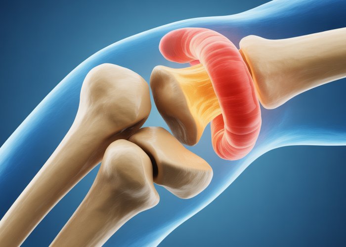
At the heart of this mechanism lies the trochlea, a spool-shaped structure on the distal end of the humerus (upper arm bone). The trochlea isn’t merely a passive element; it’s a primary weight-bearing surface, responsible for the stable and controlled movement of the elbow.
Its unique shape dictates the range of motion and contributes significantly to the joint’s overall stability.
The Elbow Joint: A Foundation for Upper Limb Dexterity
The elbow joint is more than just a connection point; it’s a facilitator of complex movements. Its primary function is to allow flexion (bending) and extension (straightening) of the arm.
This range of motion is vital for countless daily activities, from reaching for objects to performing intricate tasks. The joint also contributes to forearm rotation (pronation and supination) in conjunction with the wrist, further expanding the hand’s functional capabilities.
The elbow’s stability is paramount. Without it, even simple tasks would become difficult or impossible. This stability is achieved through a combination of bony architecture, ligamentous support, and the precise articulation of the joint surfaces, most notably the trochlea.
The Trochlea’s Critical Role in Elbow Stability and Articulation
The trochlea plays a central role in ensuring the elbow’s stability and smooth articulation. Its spool-like shape creates a secure fit with the ulna, one of the forearm bones.
This articulation is vital for resisting forces and preventing unwanted movement, particularly side-to-side instability. The trochlea’s design minimizes the risk of dislocation and allows for controlled, predictable motion.
The trochlea’s significance extends beyond stability. Its carefully contoured surface guides the movement of the ulna during flexion and extension. This allows for a smooth, almost frictionless motion, reducing wear and tear on the joint over time.
Purpose of This Article: A Comprehensive Guide
This article aims to provide a comprehensive understanding of the trochlea, a often overlooked but critically important structure of the elbow. We will delve into its anatomy, exploring its unique shape and relationship to surrounding structures.
We will also examine its biomechanical function, detailing how it enables smooth and stable elbow movement. Finally, we will discuss common injuries and conditions that can affect the trochlea.
We’ll also discuss how those injuries are diagnosed and treated, and finally discuss the rehabilitation process.
By the end of this article, you will have a complete understanding of the trochlea and its vital role in maintaining elbow health and function.
The elbow joint is more than just a connection point; it’s a facilitator of complex movements. Its primary function is to allow flexion (bending) and extension (straightening) of the arm.
This range of motion is vital for countless daily activities, from reaching for objects to performing intricate tasks. The joint also contributes to forearm rotation (pronation and supination) in conjunction with the wrist, further expanding the hand’s functional capabilities.
The elbow’s stability is paramount. Without it, even simple tasks would become difficult or impossible. This stability is achieved through a combination of bony architecture, ligamentous support, and the precise articulation of the joint surfaces, most notably the trochlea.
Anatomy: A Detailed Look at the Trochlea
Understanding the intricacies of the trochlea’s anatomy is fundamental to appreciating its functional significance and the potential consequences of injury.
The trochlea isn’t simply a rounded prominence; it’s a carefully sculpted structure whose form directly dictates the elbow’s movement capabilities. Its precise location and unique shape within the elbow joint are critical to its function.
The Trochlea and the Distal Humerus
The trochlea forms an integral part of the distal humerus, the lower end of the upper arm bone.
Specifically, it is situated on the medial aspect of the distal humerus, distinguished by its spool-like or pulley-like shape.
This shape is not arbitrary; it’s crucial for guiding the ulna during flexion and extension. The trochlea’s medial flange extends further distally than its lateral aspect.
This asymmetry contributes to the inherent stability of the elbow and guides the ulna in a slightly oblique path during movement.
Articulation with the Ulna
The trochlea’s primary articulation is with the trochlear notch of the ulna.
The trochlear notch is a concave surface that perfectly mirrors the convex shape of the trochlea.
This precise fit ensures a stable and congruent articulation. During elbow flexion and extension, the trochlear notch of the ulna glides smoothly within the trochlea’s groove.
The close congruity of these surfaces is a major factor in the elbow’s inherent stability, particularly in the sagittal plane (the plane of flexion and extension).
Relationship to Adjacent Bony Landmarks
The trochlea doesn’t exist in isolation; its function is intimately related to the surrounding bony structures of the elbow.
Understanding these relationships is crucial for appreciating the complex biomechanics of the joint.
The Capitulum
Laterally adjacent to the trochlea lies the capitulum, another articular surface on the distal humerus.
The capitulum articulates with the head of the radius.
While the trochlea primarily guides flexion and extension, the capitulum plays a more significant role in pronation and supination (rotation of the forearm).
The Epicondyles
The medial and lateral epicondyles are bony prominences located on either side of the distal humerus, proximal to the trochlea and capitulum.
These epicondyles serve as attachment points for various ligaments and muscles that contribute to elbow stability and movement.
The ulnar collateral ligament (UCL), a critical stabilizer of the elbow, originates from the medial epicondyle.
The Radial Head
The radial head, located on the proximal end of the radius, articulates with the capitulum, as mentioned above, but also has a close spatial relationship with the lateral aspect of the elbow joint.
The radial head’s ability to rotate against the capitulum is essential for forearm pronation and supination.
Understanding these relationships provides a complete picture of the trochlea’s role in elbow function.
Its precise location, unique shape, and articulation with the ulna, combined with its connection to surrounding bony landmarks, all contribute to the elbow’s remarkable range of motion and stability.
The asymmetry contributes to the inherent stability of the elbow and positions the ulna for optimal engagement with the humerus throughout the full range of motion. But how does this intricate anatomy translate into functional movement and stability?
Function and Biomechanics: How the Trochlea Enables Movement
The trochlea’s anatomical design is directly linked to its biomechanical function. It serves as the linchpin for elbow movement, specifically flexion and extension, while also contributing significantly to overall joint stability. Understanding how the trochlea facilitates these movements is crucial for appreciating its importance in upper limb function.
Flexion and Extension: The Trochlea’s Primary Role
The trochlea’s primary role is to allow flexion (bending) and extension (straightening) of the elbow joint.
This fundamental movement is essential for a wide range of daily activities, from lifting objects to performing delicate hand movements.
The ulna, specifically the trochlear notch, articulates with the trochlea in a hinge-like fashion. This articulation allows the forearm to move towards (flexion) and away from (extension) the upper arm. The smooth, curved surface of the trochlea ensures a fluid and consistent range of motion during these movements.
The Trochlea’s Shape: A Key to Smooth and Controlled Motion
The trochlea’s unique spool-like shape is not merely a structural feature; it is integral to the smoothness and control of elbow movement.
This shape allows for a congruent articulation with the ulna’s trochlear notch, ensuring that the joint surfaces remain in constant contact throughout the full range of motion. This congruency minimizes stress concentration and promotes even distribution of forces across the joint.
The medial flange of the trochlea, which extends further distally, acts as a guiding rail for the ulna during flexion and extension.
This anatomical feature prevents excessive side-to-side movement and contributes to the inherent stability of the elbow joint.
Ligamentous Support: UCL and RCL’s Contribution to Stability
While the trochlea’s bony architecture provides inherent stability, the ligaments surrounding the elbow joint play a crucial role in reinforcing this stability, especially during movement.
The two primary ligaments involved are the ulnar collateral ligament (UCL) and the radial collateral ligament (RCL).
Ulnar Collateral Ligament (UCL)
The UCL is located on the medial side of the elbow and is the primary stabilizer against valgus stress (stress applied to the outside of the elbow, causing it to bend inward). It prevents excessive abduction of the forearm.
This ligament is particularly important during overhead activities, such as throwing, where significant valgus forces are generated.
Radial Collateral Ligament (RCL)
The RCL is located on the lateral side of the elbow and provides stability against varus stress (stress applied to the inside of the elbow, causing it to bend outward).
It also contributes to overall elbow stability by resisting rotational forces.
Both the UCL and RCL work in concert with the trochlea’s bony architecture to maintain a stable and functional elbow joint throughout its range of motion. Injuries to these ligaments, particularly the UCL, can significantly compromise elbow stability and function, often requiring medical intervention.
The trochlea’s Shape: A Key to Smooth and Controlled Motion
The trochlea’s unique spool-like shape is not merely a structural feature; it is integral to the smoothness and control of elbow movement. This shape allows for a congruent articulation with the ulna’s trochlear notch, ensuring that the joint surfaces remain in constant contact throughout the full range of motion. This congruency minimizes stress concentration and enhances stability. But what happens when this carefully orchestrated anatomy is disrupted by injury or disease?
Common Injuries and Conditions Affecting the Trochlea
The trochlea, though robust in design, is susceptible to various injuries and conditions that can compromise elbow function. These issues range from traumatic events like dislocations and fractures to degenerative processes such as osteoarthritis. Understanding these conditions is crucial for effective diagnosis and treatment.
Elbow Dislocation
Elbow dislocations are significant injuries that can severely impact the trochlea and overall joint stability. These dislocations typically occur due to high-energy trauma, such as falls onto an outstretched arm.
When the elbow dislocates, the ulna is forced out of its normal articulation with the humerus, directly affecting the trochlea’s alignment. This misalignment can lead to significant instability and damage to the surrounding ligaments and soft tissues.
The severity of the dislocation can range from simple, where only the joint is disrupted, to complex, involving associated fractures.
Symptoms and Diagnosis
The symptoms of an elbow dislocation are usually immediate and pronounced:
- Severe pain
- Obvious deformity of the elbow
- Inability to move the arm
Diagnosis typically involves a physical examination by a medical professional, followed by imaging studies. X-rays are essential to confirm the dislocation and identify any associated fractures. In some cases, an MRI may be necessary to assess ligament damage.
Treatment Approaches
The primary goal of treatment for an elbow dislocation is to reduce the dislocation, meaning to relocate the bones back into their proper alignment. This is typically done under sedation or anesthesia to minimize pain and muscle spasm.
Following reduction, the elbow is usually immobilized in a splint or cast for a period to allow the ligaments to heal. Physical therapy is a crucial component of rehabilitation, focusing on restoring range of motion, strength, and stability to the elbow.
In cases of complex dislocations with associated fractures or significant ligament damage, surgery may be required to repair the damaged structures and ensure long-term stability.
Fractures Involving the Trochlea
Fractures that directly involve the trochlea are complex injuries that can have significant implications for elbow function. These fractures can occur due to direct trauma to the elbow or from forces transmitted through the forearm.
The capitellum and trochlea are particularly vulnerable to injury in the setting of distal humerus fractures.
Types of Fractures
Several types of fractures can involve the trochlea, including:
- Capitellum Fractures: Though primarily involving the capitellum, these fractures can extend into the trochlea.
- Trochlear Groove Fractures: Direct injuries to the trochlea.
- Distal Humerus Fractures: Complex fractures that involve both the trochlea and capitellum, as well as the humeral condyles.
Causes, Diagnosis, and Management
Trochlear fractures are commonly caused by high-impact trauma.
Diagnosis typically involves X-rays to visualize the fracture pattern. CT scans may be used to provide more detailed information about the fracture and guide surgical planning.
Management of trochlea fractures depends on the severity and displacement of the fracture. Non-displaced fractures may be treated with immobilization in a cast or splint, followed by physical therapy.
Displaced fractures usually require surgical intervention to restore the joint surface and ensure proper alignment. Surgical options may include open reduction and internal fixation (ORIF) with plates and screws or, in severe cases, elbow replacement.
Osteoarthritis of the Elbow
Osteoarthritis (OA) is a degenerative joint disease that can affect the elbow, leading to pain, stiffness, and decreased function. OA involves the progressive breakdown of cartilage within the joint.
While osteoarthritis can affect any part of the elbow, the trochlea is particularly vulnerable due to its role as a major weight-bearing surface. As the cartilage wears away, the underlying bone is exposed, leading to pain and inflammation.
Impact on the Trochlea
The degradation of cartilage on the trochlea results in bone-on-bone contact, causing pain and limiting the smooth gliding motion of the elbow joint.
Over time, this can lead to the formation of bone spurs (osteophytes) around the joint, further restricting movement and causing pain.
Symptoms and Management
The symptoms of elbow osteoarthritis typically develop gradually and include:
- Pain, especially with activity
- Stiffness, particularly in the morning or after periods of inactivity
- Swelling around the elbow joint
- Decreased range of motion
- Grinding or clicking sensation in the elbow
Management of elbow osteoarthritis aims to relieve pain, improve function, and slow the progression of the disease.
Non-surgical treatments include:
- Pain relievers, such as NSAIDs or acetaminophen
- Physical therapy to improve range of motion and strength
- Corticosteroid injections to reduce inflammation
- Viscosupplementation injections to lubricate the joint
In severe cases where non-surgical treatments are ineffective, surgery may be considered. Surgical options include:
- Arthroscopic debridement to remove bone spurs and loose cartilage
- Elbow replacement (arthroplasty) for end-stage arthritis.
Diagnosis and Imaging: Seeing the Trochlea
Given the trochlea’s pivotal role in elbow function, accurately diagnosing any related issues is paramount for effective treatment and restoring optimal joint mechanics. A misdiagnosis or delayed diagnosis can lead to chronic pain, instability, and impaired range of motion. Fortunately, advancements in imaging technology provide clinicians with powerful tools to visualize the trochlea and surrounding structures in detail, allowing for a more precise assessment of the underlying problem.
The Necessity of a Precise Diagnosis
Effective management of trochlea-related problems hinges on accurate diagnosis. The complexity of the elbow joint, with its intricate network of bones, ligaments, and tendons, requires a comprehensive evaluation to pinpoint the source of pain and dysfunction.
A precise diagnosis informs treatment decisions, whether conservative measures like physical therapy or surgical interventions are required. Furthermore, it allows healthcare professionals to tailor treatment plans to address the specific nature and extent of the trochlea’s involvement, maximizing the chances of successful recovery and long-term joint health.
X-ray: Visualizing Bony Architecture
X-rays, or radiographs, remain a fundamental diagnostic tool for evaluating the trochlea and the overall bony architecture of the elbow. X-rays use electromagnetic radiation to create images of the skeletal structures within the body. Because dense materials like bone block more radiation than soft tissue, bones appear white on an X-ray image, allowing easy evaluation.
X-rays are particularly effective in identifying fractures, dislocations, and other bony abnormalities affecting the trochlea. They can reveal the presence of fracture lines, the displacement of bone fragments, and the overall alignment of the elbow joint.
In cases of suspected trauma or acute injury, X-rays are typically the first-line imaging modality employed to assess the integrity of the trochlea and rule out any significant bony injuries. Weight-bearing X-rays can also be performed to assess joint space narrowing and alignment under load, which can be indicative of osteoarthritis or other degenerative conditions.
MRI (Magnetic Resonance Imaging): Delving into Soft Tissues
While X-rays excel at visualizing bony structures, Magnetic Resonance Imaging (MRI) provides a more detailed assessment of the soft tissues surrounding the trochlea, including ligaments, tendons, cartilage, and muscles. MRI uses strong magnetic fields and radio waves to generate cross-sectional images of the body.
MRI is particularly valuable in identifying subtle injuries that may not be apparent on X-rays, such as ligament tears, cartilage damage, or bone bruises (bone marrow edema). It can also detect early signs of osteoarthritis, such as cartilage thinning or joint effusion (fluid accumulation).
The ability of MRI to visualize soft tissue structures with high resolution makes it an indispensable tool for diagnosing a wide range of trochlea-related conditions, including:
- Ulnar collateral ligament (UCL) injuries.
- Radial collateral ligament (RCL) injuries.
- Osteochondral lesions (damage to cartilage and underlying bone).
- Soft tissue impingement.
- Occult fractures (stress fractures not visible on X-ray).
By providing a comprehensive view of both bony and soft tissue structures, MRI plays a crucial role in guiding treatment decisions and optimizing patient outcomes. In many cases, MRI findings can help to differentiate between various potential diagnoses and determine the most appropriate course of action.
Treatment Options: Restoring Elbow Function
Having pinpointed the issue affecting the trochlea and its surrounding structures, the next crucial step involves formulating an effective treatment plan. Fortunately, a range of treatment options exist, from conservative non-surgical approaches to advanced surgical interventions, designed to alleviate pain, restore function, and improve overall elbow health.
Non-Surgical Treatment: A Conservative Approach
In many cases, particularly for less severe injuries or conditions, non-surgical treatment options can be highly effective in managing trochlea-related problems. These approaches prioritize pain relief, inflammation reduction, and the restoration of strength and range of motion.
Physical Therapy: The Cornerstone of Rehabilitation
Physical therapy plays a central role in non-surgical management. A skilled physical therapist will develop an individualized program tailored to the specific needs and limitations of the patient.
This program will typically include a combination of:
- Range-of-motion exercises: These exercises aim to gently restore the full range of movement in the elbow joint, preventing stiffness and promoting joint lubrication.
- Strengthening exercises: Targeting the muscles surrounding the elbow, such as the biceps, triceps, and forearm muscles, helps to stabilize the joint and improve its ability to withstand stress.
- Proprioceptive exercises: These exercises focus on improving the patient’s awareness of their elbow’s position in space, enhancing coordination and preventing re-injury.
- Manual therapy techniques: Hands-on techniques, such as joint mobilization and soft tissue massage, can help to reduce pain, improve joint mechanics, and release muscle tension.
Pain Management and Supportive Care
Alongside physical therapy, various pain management strategies and supportive care options can provide significant relief. These may include:
- Pain medication: Over-the-counter pain relievers, such as acetaminophen or ibuprofen, can help to reduce mild to moderate pain. In some cases, a physician may prescribe stronger pain medications.
- Ice and heat therapy: Applying ice packs to the elbow can help to reduce inflammation and pain, while heat therapy can soothe muscles and improve circulation.
- Bracing or splinting: A brace or splint can provide support and stability to the elbow joint, reducing stress on the trochlea and surrounding structures.
- Injections: Corticosteroid injections can be administered to reduce inflammation and pain within the elbow joint. However, these injections are typically used sparingly due to potential long-term side effects.
Surgical Intervention: When is it Necessary?
While non-surgical treatments are often successful, surgery may be necessary in certain situations. These include:
- Severe fractures involving the trochlea: If the trochlea is severely fractured, surgery may be required to realign the bone fragments and stabilize the joint.
- Unstable elbow dislocations: In cases of recurrent or unstable elbow dislocations, surgery may be needed to repair damaged ligaments and restore joint stability.
- End-stage osteoarthritis: When osteoarthritis has severely damaged the cartilage in the elbow joint, leading to significant pain and loss of function, joint replacement surgery may be considered.
The Role of the Orthopedic Surgeon
An orthopedic surgeon, specializing in treating musculoskeletal conditions, plays a crucial role in determining whether surgery is the appropriate course of action. They will carefully evaluate the patient’s condition, considering the severity of the injury, the patient’s age and activity level, and the response to non-surgical treatments.
If surgery is deemed necessary, the orthopedic surgeon will perform the procedure, utilizing advanced techniques and technologies to restore elbow function and alleviate pain. The specific surgical approach will vary depending on the underlying problem. It can involve procedures such as:
- Open Reduction and Internal Fixation (ORIF): A surgery to repair severely fractured trochlea, which requires the surgeon to make an incision to access the bone fragments. The fragments are then repositioned (reduced) into their normal alignment and held together with screws, plates, or wires (internal fixation).
- Ligament Reconstruction: This procedure involves repairing or reconstructing damaged ligaments to stabilize the elbow joint and prevent further dislocations.
- Elbow Arthroplasty (Joint Replacement): In this surgery, the damaged joint surfaces are replaced with artificial components.
Alongside medical and surgical interventions, a comprehensive approach to recovery necessitates a focus on restoring the elbow’s functionality, strength, and range of motion. This is where the crucial role of a structured rehabilitation program comes into play, bridging the gap between treatment and a return to normal activities.
Rehabilitation and Recovery: Getting Back to Normal
The journey to regaining full elbow function after a trochlea-related injury or surgery extends far beyond the initial treatment phase. A well-designed and diligently followed rehabilitation program is the cornerstone of a successful recovery. It’s not merely about healing; it’s about rebuilding strength, restoring flexibility, and regaining confidence in using the elbow.
The Importance of a Structured Program
Adhering to a structured rehabilitation program is not optional; it’s essential. It ensures that the healing tissues are progressively loaded, preventing stiffness, minimizing the risk of re-injury, and optimizing the long-term outcome. Think of it as a carefully orchestrated symphony, where each exercise and activity plays a specific role in the overall healing process. Deviating from this plan can disrupt the harmony and hinder progress.
A qualified physical therapist will tailor the program to your specific needs, considering the nature and severity of the injury, your overall health, and your individual goals.
Exercises and Activities for Restoring Function
Rehabilitation programs typically incorporate a variety of exercises and activities to address different aspects of elbow function:
-
Range-of-Motion Exercises: These gentle movements aim to restore the full spectrum of elbow flexion, extension, pronation (turning the palm down), and supination (turning the palm up). Examples include:
- Active assisted range of motion, where you use your other arm to gently guide the movement.
- Passive range of motion, where the therapist moves your arm for you.
-
Strengthening Exercises: Once pain and inflammation subside, the focus shifts to rebuilding strength in the muscles surrounding the elbow. This includes exercises for the biceps, triceps, forearm muscles, and shoulder muscles. Examples include:
- Isometric exercises, where you contract the muscles without moving the joint.
- Theraband exercises, using resistance bands to provide progressive challenge.
- Light weight training, gradually increasing the weight as tolerated.
-
Proprioceptive Exercises: These exercises improve your awareness of your elbow’s position in space, enhancing coordination and stability. Examples include:
- Balance exercises on unstable surfaces.
- Reaching and grasping activities with varying speeds and loads.
-
Functional Activities: As strength and range of motion improve, the program will incorporate activities that mimic real-life movements, such as lifting, carrying, and throwing. The goal is to gradually return you to your pre-injury level of function.
Recovery Timeline and Individual Variability
The recovery timeline after a trochlea-related injury or surgery is highly variable and depends on several factors:
- Severity of the injury
- Type of treatment received
- Individual healing capacity
- Adherence to the rehabilitation program
A general timeline can be provided, but it’s crucial to understand that progress may not be linear. Some days will be better than others, and setbacks are a normal part of the recovery process. Patience, persistence, and close communication with your physical therapist are key.
In general, expect the initial phase of recovery, focusing on pain management and early range of motion, to last for several weeks. Strengthening and functional activities are gradually introduced over the following months. A full return to activity, including sports or heavy labor, may take several months to a year.
Remember, the recovery journey is a marathon, not a sprint. It requires dedication, commitment, and a collaborative approach between you, your physical therapist, and your physician. By actively participating in your rehabilitation program and staying positive, you can maximize your chances of a successful outcome and a return to a pain-free, functional elbow.
Trochlea of Elbow FAQs
Here are some frequently asked questions about the trochlea of the elbow, aiming to clarify common points and provide helpful information.
Where exactly is the trochlea of elbow located?
The trochlea is located at the distal end of the humerus, which is the long bone in your upper arm. It’s on the medial (inner) side of the elbow joint, articulating with the ulna.
What is the trochlea of elbow’s main function?
Its primary function is to provide a smooth, stable surface for the ulna to articulate with, enabling flexion and extension movements of the elbow joint. This allows you to bend and straighten your arm.
How does an injury to the trochlea of elbow affect elbow movement?
Damage or injury to the trochlea can significantly restrict the elbow’s range of motion. Fractures or cartilage damage can cause pain, stiffness, and instability, making it difficult to perform everyday tasks.
What are the common treatments for trochlea of elbow injuries?
Treatment options vary depending on the severity of the injury. They may include conservative methods like rest, ice, compression, and elevation (RICE), pain medication, and physical therapy. In severe cases, surgery might be necessary to repair the trochlea of elbow or restore joint stability.
So, there you have it – everything you need to know about the trochlea of elbow! Hopefully, you found this helpful. Now you can impress your friends with your knowledge of elbow anatomy! Keep learning, and take care of those elbows!