The intricate world of transversal cut anatomy, often explored through advanced imaging techniques like MRI, offers critical insights into anatomical structures. Detailed understanding of transversal cut anatomy is essential for professionals in fields such as radiology. Medical schools, including institutions prominent in surgical training, emphasize transversal cut anatomy. The anatomical precision demonstrated in classic texts like Gray’s Anatomy is mirrored in contemporary applications of transversal cut anatomy, underlining its enduring relevance for medical practice.
Crafting the Ultimate "Transversal Cut Anatomy" Visual Guide: Article Layout
To create a comprehensive and engaging visual guide on transversal cut anatomy, a well-structured layout is essential. The focus must always be on clarity and ease of understanding, particularly for readers who may be new to the subject. The following layout provides a framework for presenting this complex topic in an accessible manner.
I. Introduction: Defining Transversal Cut Anatomy
- Purpose: To establish the importance and relevance of understanding transversal cut anatomy.
- Content: Start by clearly defining what a transversal cut (also known as axial or horizontal cut) is in anatomical terms.
- Explain that it’s a plane that divides the body or an organ into superior (upper) and inferior (lower) parts.
- Briefly explain the significance of using this plane in medical imaging (CT scans, MRI).
- Visuals: Include an introductory image. This could be:
- A diagram showing the three main anatomical planes (sagittal, coronal, and transversal) with the transversal plane highlighted.
- A simplified illustration of a human body with a transversal cut indicated, showing the resulting upper and lower sections.
- Example phrasing: "Transversal cut anatomy is fundamental to interpreting medical images and understanding the internal structure of the human body. This guide will provide a comprehensive visual exploration of anatomical structures as they appear in transversal sections."
II. Key Anatomical Terminology for Orientation
- Purpose: To ensure a common understanding of anatomical terms used throughout the guide.
- Content: Define key terms related to position and direction, as these are critical for understanding transversal sections.
- Superior vs. Inferior
- Anterior (Ventral) vs. Posterior (Dorsal)
- Medial vs. Lateral
- Proximal vs. Distal (if applicable to limb sections)
- Right vs. Left
- Visuals:
- Diagram illustrating these terms on a human figure. Use arrows and clear labels.
-
Table Example:
Term Definition Superior Above or towards the head Inferior Below or towards the feet Anterior Towards the front of the body Posterior Towards the back of the body Medial Towards the midline of the body Lateral Away from the midline of the body
III. Transversal Anatomy of Major Body Regions
- Purpose: To systematically explore transversal anatomy across different regions of the body.
-
Structure: Divide this section into subsections based on major body regions.
A. Transversal Anatomy of the Head
- Content: Describe the key structures visible in a transversal cut of the head.
- Brain (cerebrum, cerebellum, brainstem)
- Skull bones
- Sinuses
- Eyes
- Important blood vessels (e.g., carotid arteries)
- Visuals: Use labeled images of transversal CT or MRI scans of the head. Highlight specific structures with arrows and annotations. Each structure should be briefly described in the caption.
B. Transversal Anatomy of the Neck
- Content: Outline the key structures visible in a transversal cut of the neck.
- Vertebrae
- Spinal cord
- Esophagus
- Trachea
- Thyroid gland
- Major blood vessels (e.g., carotid arteries, jugular veins)
- Muscles
- Visuals: Use labeled images of transversal CT or MRI scans of the neck. Label each visible component.
C. Transversal Anatomy of the Thorax (Chest)
- Content: Describe the key structures visible in a transversal cut of the thorax. This should be broken down further.
- Lungs
- Heart
- Ribs
- Vertebrae
- Esophagus
- Trachea/Bronchi
- Major blood vessels (aorta, vena cava, pulmonary arteries/veins)
- Muscles
- Mediastinum (highlighting its contents)
- Visuals: Labeled CT/MRI images of the thorax at different levels (e.g., at the level of the heart, at the level of the aortic arch).
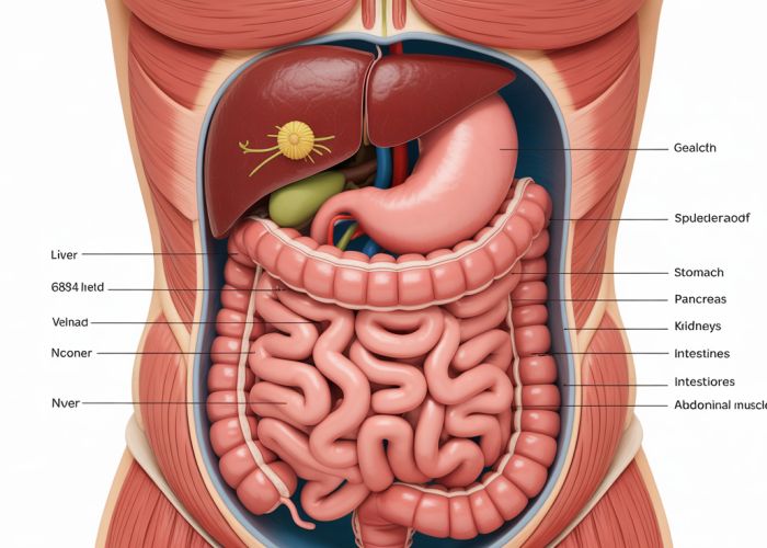
D. Transversal Anatomy of the Abdomen
- Content: Detail the key structures visible in a transversal cut of the abdomen. This also needs further breakdown.
- Liver
- Spleen
- Stomach
- Pancreas
- Kidneys
- Intestines (small and large)
- Vertebrae
- Aorta/Inferior Vena Cava
- Abdominal muscles
- Visuals: Labeled CT/MRI images of the abdomen at different levels to demonstrate varying anatomical relationships.
E. Transversal Anatomy of the Pelvis
- Content: Explain the key structures visible in a transversal cut of the pelvis.
- Pelvic bones
- Rectum
- Bladder
- Reproductive organs (uterus/ovaries in females, prostate/seminal vesicles in males)
- Major blood vessels
- Pelvic muscles
- Visuals: Labeled CT/MRI images of the pelvis, highlighting the differences between male and female anatomy.
F. Transversal Anatomy of the Limbs (Upper and Lower)
- Content: Describe the key structures visible in transversal cuts of the upper and lower limbs. This should be generalized, as specific levels (e.g., mid-thigh, mid-arm) would require their own, more detailed sections.
- Bones (humerus, femur, radius, ulna, tibia, fibula)
- Muscles (major muscle groups)
- Blood vessels (major arteries and veins)
- Nerves (major nerve trunks)
- Visuals: Diagrams or simplified illustrations showing the arrangement of muscles, bones, vessels, and nerves in a typical limb cross-section.
- Content: Describe the key structures visible in a transversal cut of the head.
IV. Clinical Significance of Transversal Anatomy
- Purpose: To highlight the real-world applications of understanding transversal cut anatomy in a medical context.
- Content: Provide brief examples of how transversal imaging (CT, MRI) is used to diagnose and monitor various conditions.
- Example 1: Detecting brain tumors on CT scans.
- Example 2: Identifying abdominal aortic aneurysms.
- Example 3: Diagnosing fractures.
- Example 4: Assessing organ size and shape (e.g., liver enlargement).
- Visuals: Include simplified examples of clinical images demonstrating pathological conditions in transversal sections. Be mindful of privacy and use appropriately sourced, publicly available images or diagrams. A caption explaining what the image shows is essential.
- Ethical Note: Emphasize that interpreting medical images requires expertise and should only be done by qualified healthcare professionals. This guide is for educational purposes only.
This detailed layout ensures that the "Transversal Cut Anatomy" visual guide is well-structured, informative, and accessible to a broad audience.
Transversal Cut Anatomy: Frequently Asked Questions
Here are some common questions readers have about understanding transversal cut anatomy after reviewing our visual guide. We hope these answers clarify any points.
What exactly is a transversal cut in anatomy?
A transversal cut, also known as an axial or horizontal cut, divides the body or organ into superior (upper) and inferior (lower) parts. Understanding this plane is fundamental to interpreting medical imaging like CT scans. It’s a key view when studying transversal cut anatomy.
Why is understanding transversal cut anatomy important?
It’s crucial for interpreting medical images like CT and MRI scans. These scans are frequently displayed as a series of transversal slices. Doctors use their knowledge of transversal cut anatomy to diagnose various conditions.
How does transversal cut anatomy differ from sagittal or coronal anatomy?
Sagittal cuts divide the body into left and right portions. Coronal cuts divide it into front (anterior) and back (posterior) portions. Transversal cuts are perpendicular to both, providing a different perspective of internal structures. The combination of all three planes offers a comprehensive view of anatomy.
What are some common anatomical landmarks identified in a transversal cut?
Depending on the level of the cut, you might see the vertebrae, major blood vessels (aorta, vena cava), organs like the liver, spleen, and kidneys, and various muscle groups. Familiarity with these landmarks is essential for correctly interpreting transversal cut anatomy images.
And that’s a wrap on our deep dive into transversal cut anatomy! Hopefully, this visual guide made understanding these complex images a little easier. Now go forth and impress your colleagues with your newfound knowledge! See you in the next article!