The process of gene expression fundamentally depends on transcription, a crucial stage visually represented by transcription diagram biology. These diagrams, often explored using tools like BioRender, offer a streamlined way to understand how genetic information flows. Understanding transcription is core for any research taking place within organizations such as the National Institutes of Health (NIH). This article simplifies the interpretation of transcription diagrams, unlocking a deeper understanding of biological processes and allowing students and researcher alike to easily see the interactions that make up the process of transcription in biology.
At the heart of molecular biology lies a fundamental principle, a cornerstone upon which our understanding of life is built: the central dogma. This elegant concept describes the flow of genetic information within a biological system, from DNA to RNA to protein. Understanding this flow is crucial to deciphering the mysteries of life itself.
The Central Dogma: DNA → RNA → Protein
The central dogma, often represented as DNA → RNA → Protein, outlines the process by which the instructions encoded in our DNA are ultimately used to create the proteins that carry out virtually every function in our cells.
DNA, the blueprint of life, contains the genetic code.
RNA serves as an intermediary, carrying the instructions from DNA to the protein-building machinery.
Proteins, the workhorses of the cell, perform a vast array of tasks, from catalyzing biochemical reactions to providing structural support.
Transcription: The First Step in Gene Expression
Transcription is the vital first step in gene expression, the process by which the information encoded in a gene is used to synthesize a functional gene product (protein or RNA).
It’s the process of creating a RNA copy from a DNA template. Think of it as making a photocopy of a specific instruction manual page (DNA) so the information can be brought to where proteins are built.
This process ensures that the correct proteins are produced at the right time and in the right amounts. Without transcription, the genetic information stored in DNA would be inaccessible, rendering the cell unable to function.
Why Transcription Diagrams Matter
Transcription diagrams are visual representations of this crucial process. They provide a simplified yet comprehensive overview, allowing us to understand the complex molecular interactions that drive transcription.
For biology students, transcription diagrams offer a powerful tool for visualizing and internalizing the key concepts. They bridge the gap between abstract ideas and concrete understanding.
For researchers, these diagrams serve as valuable aids in designing experiments, interpreting results, and communicating their findings to others.
What We Will Cover
In this exploration, we will delve into the intricacies of transcription diagrams, uncovering their secrets and empowering you to interpret them with confidence.
We will explore the players involved, the steps of the process, and the variations that exist in different organisms.
Prepare to embark on a journey that will unlock a deeper understanding of one of the most fundamental processes in biology.
Transcription Demystified: The Core Process Explained
Transcription, at its core, is the mechanism by which the genetic information encoded within DNA is copied into RNA.
But why is this copying necessary? DNA, the master blueprint, resides safely within the nucleus.
To utilize the information it holds for protein synthesis, a mobile intermediary is required – RNA.
This process, therefore, allows the genetic instructions to be transported from the nucleus to the ribosomes in the cytoplasm, where proteins are synthesized. It is the essential first step in gene expression.
The Key Players in Transcription
Several key molecules orchestrate the intricate process of transcription. Let’s take a closer look at each:
-
DNA (The Template): DNA serves as the original source of genetic information. Specific regions of DNA, called genes, are used as templates to synthesize complementary RNA molecules. Only one strand of the DNA, the template strand, is used for transcription.
-
RNA (The Product): RNA is the resulting molecule that carries the genetic information copied from DNA. Unlike DNA, RNA is typically single-stranded.
Furthermore, RNA uses uracil (U) instead of thymine (T) as one of its nucleotide bases. Multiple types of RNA exist, each playing a distinct role in the cell. However, mRNA (messenger RNA) is the primary product of transcription that carries the genetic code for protein synthesis.
-
RNA Polymerase (The Enzyme): RNA polymerase is the central enzyme that catalyzes the synthesis of RNA.
It binds to DNA, unwinds the double helix locally, and reads the template strand to synthesize a complementary RNA molecule.
RNA polymerase moves along the DNA template, adding RNA nucleotides one by one to the growing RNA strand.
-
Transcription Factors (The Regulators): Transcription factors are proteins that regulate the activity of RNA polymerase. They can either enhance or repress transcription, controlling when and how much of a gene is expressed.
Some transcription factors bind directly to DNA, while others interact with RNA polymerase or other transcription factors.
These factors play a crucial role in ensuring that genes are expressed at the right time and in the right cells.
The Promoter Region: Where Transcription Begins
The initiation of transcription doesn’t happen just anywhere on the DNA. It starts at a specific region called the promoter.
The promoter is a DNA sequence located upstream of a gene that signals the start of transcription. It acts as a binding site for RNA polymerase and transcription factors.
One crucial element within many promoters is the TATA box.
The TATA box is a DNA sequence rich in thymine (T) and adenine (A) bases. It serves as a recognition site for transcription factors.
These factors help to position RNA polymerase correctly on the DNA.
Once RNA polymerase is properly positioned at the promoter, transcription can begin. The TATA box is thus a critical landmark that dictates where and when transcription will occur.
Decoding Transcription Diagrams: A Step-by-Step Guide
Having examined the fundamental process of transcription and the molecules that drive it, we now turn our attention to interpreting the visual language used to represent this complex event. Transcription diagrams are essential tools for visualizing and understanding the flow of genetic information, but deciphering them requires familiarity with their conventions and components.
Understanding DNA Strands: Template vs. Coding
At the heart of a transcription diagram lies the DNA double helix. It is critical to distinguish between the two strands: the template strand (also known as the non-coding strand) and the coding strand (also known as the non-template strand).
The template strand is the DNA strand that RNA polymerase uses as a guide to synthesize the RNA molecule. Its sequence is complementary to the RNA sequence that is produced (with uracil replacing thymine).
The coding strand, on the other hand, has the same sequence as the RNA molecule (again, with uracil replacing thymine). It is called the "coding strand" because its sequence corresponds to the codons that are eventually translated into amino acids to build a protein.
Understanding the distinction between these strands is crucial for accurately interpreting which part of the DNA is being transcribed and what the resulting RNA sequence will be. The template strand serves as the blueprint, while the coding strand provides a direct representation of the final RNA product.
The Three Stages of Transcription: A Visual Walkthrough
Transcription diagrams often depict the process as a series of stages: initiation, elongation, and termination. Each stage has distinct characteristics that are visually represented in the diagram.
Initiation: Starting the Process
Initiation is the beginning of transcription, where RNA polymerase binds to a specific region of DNA called the promoter. This promoter region signals the start of a gene.
Often, transcription factors, proteins that help regulate gene expression, assist RNA polymerase in binding to the promoter. The diagram will likely show the RNA polymerase bound to the DNA near the promoter region, sometimes with transcription factors nearby. A key sequence within the promoter is often the TATA box, vital for positioning RNA polymerase.
Elongation: Building the RNA Molecule
Once RNA polymerase is bound to the promoter, elongation begins. During elongation, RNA polymerase moves along the template strand of the DNA. It reads the DNA sequence and adds complementary RNA nucleotides to the growing RNA molecule.
The diagram will show RNA polymerase moving along the DNA, with the newly synthesized RNA molecule trailing behind it. The direction of movement is important: the arrow typically indicates the 5′ to 3′ direction of RNA synthesis.
Termination: Ending the Process
Termination is the final stage, where transcription stops. RNA polymerase reaches a specific sequence called the terminator sequence on the DNA.
This sequence signals RNA polymerase to detach from the DNA and release the newly synthesized RNA molecule. The diagram will show RNA polymerase detaching from the DNA at the terminator sequence, with the complete RNA molecule now free.
Visualizing RNA Polymerase Movement
A well-constructed transcription diagram clearly illustrates the movement of RNA polymerase along the DNA template. The diagram effectively conveys the direction of transcription, the addition of nucleotides, and the progression of the enzyme.
This visual representation makes it easier to understand the dynamic nature of transcription and how the RNA molecule is assembled step-by-step. By carefully examining the diagram, one can gain a deeper appreciation for the intricate choreography of molecules involved in this fundamental biological process.
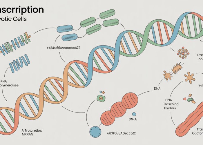
Having navigated the intricacies of transcription diagrams and established a firm understanding of the core transcriptional process, it’s time to address a critical distinction: the differences between transcription as it occurs in prokaryotic and eukaryotic cells. While the fundamental principles remain the same, the context and complexity vary significantly, particularly regarding cellular location and post-transcriptional modifications.
Prokaryotic vs. Eukaryotic Transcription: Key Differences
The process of transcription, while fundamentally similar across all life forms, exhibits key differences between prokaryotes and eukaryotes. These differences stem from variations in cellular structure, regulatory mechanisms, and post-transcriptional processing. Understanding these distinctions is crucial for a comprehensive understanding of gene expression.
Prokaryotic Simplicity: Transcription in the Cytoplasm
In prokaryotic cells, such as bacteria and archaea, transcription occurs in the cytoplasm. This is because prokaryotes lack a membrane-bound nucleus. The absence of a nucleus means that transcription and translation are spatially and temporally coupled.
As the mRNA molecule is being transcribed from the DNA template, ribosomes can immediately bind to it and begin translating it into protein. This direct link between transcription and translation allows for rapid gene expression in response to environmental changes.
The simpler cellular structure of prokaryotes also means that the regulatory mechanisms governing transcription are generally less complex than those found in eukaryotes.
Eukaryotic Complexity: Transcription in the Nucleus and Beyond
Eukaryotic transcription, on the other hand, is a far more complex process that occurs within the nucleus. The presence of a nuclear membrane separates transcription from translation, adding an additional layer of regulation.
The Role of the Nucleus
The nucleus provides a specialized environment for transcription, protecting the DNA from damage and allowing for the precise regulation of gene expression. It also allows for post-transcriptional modifications that are essential for mRNA maturation and function.
Post-Transcriptional Modifications: A Hallmark of Eukaryotic Transcription
One of the key differences between prokaryotic and eukaryotic transcription is the extensive post-transcriptional modification of mRNA in eukaryotes. These modifications include:
-
5′ Capping: The addition of a modified guanine nucleotide to the 5′ end of the mRNA molecule, protecting it from degradation and enhancing translation.
-
Splicing: The removal of non-coding regions called introns from the pre-mRNA molecule, and the joining of coding regions called exons to form the mature mRNA.
-
3′ Polyadenylation: The addition of a tail of adenine nucleotides (the poly(A) tail) to the 3′ end of the mRNA molecule, which also protects it from degradation and enhances translation.
Splicing is a particularly important process in eukaryotes, allowing for a single gene to encode multiple different proteins through alternative splicing mechanisms. This dramatically increases the protein diversity that can be generated from a limited number of genes.
These post-transcriptional modifications are essential for the stability, translatability, and ultimately, the function of the mRNA molecule in eukaryotes. They are also targets for regulatory mechanisms that control gene expression.
In summary, while the fundamental principles of transcription are conserved across prokaryotes and eukaryotes, the process is significantly more complex in eukaryotes due to the presence of a nucleus and the extensive post-transcriptional modification of mRNA. These differences reflect the greater complexity of eukaryotic cells and the need for more sophisticated regulatory mechanisms to control gene expression.
From Transcription to mRNA: The Journey of the Messenger
Having navigated the intricacies of transcription diagrams and established a firm understanding of the core transcriptional process, it’s time to address a critical distinction: the differences between transcription as it occurs in prokaryotic and eukaryotic cells. While the fundamental principles remain the same, the context and complexity vary significantly, particularly regarding cellular location and post-transcriptional modifications.
Transcription serves as the initial step in gene expression. But the RNA transcript produced isn’t the final product. In fact, the RNA molecule needs to embark on a journey to fulfill its role in protein synthesis. This journey culminates in the production of messenger RNA (mRNA). mRNA carries the genetic code to the ribosomes. Here, the code is translated into the amino acid sequence of a protein.
mRNA: The Coded Dispatch
mRNA’s primary function is to serve as the intermediary between the genetic information stored in DNA and the protein synthesis machinery of the cell.
Think of DNA as the master blueprint locked away in the nucleus. mRNA is a carefully copied and deliverable version of that blueprint. This process is crucial because DNA cannot directly participate in protein synthesis. The genetic code, written in the sequence of nucleotide bases (adenine, guanine, cytosine, and uracil), is carried by mRNA. It is then decoded by ribosomes to assemble proteins.
In eukaryotes, the journey of mRNA is a carefully orchestrated process. The mRNA must undergo processing and transport before it can be translated.
Untranslated Regions: More Than Just Spacers
mRNA molecules contain regions that are not directly translated into protein. These are known as the untranslated regions (UTRs). Specifically, the 5′ UTR and 3′ UTR flank the protein-coding region. While not coding for amino acids, these regions play vital roles in regulating mRNA translation and stability.
The 5′ UTR: Guiding Ribosome Binding
The 5′ UTR is located at the beginning of the mRNA molecule. This is the region where the ribosome initially binds to the mRNA. This region often contains regulatory elements that influence the efficiency of translation initiation.
The length and sequence of the 5′ UTR can affect how easily the ribosome can bind and begin scanning for the start codon (usually AUG).
Certain sequences within the 5′ UTR can form secondary structures. These structures can either enhance or inhibit ribosome binding, thereby modulating protein synthesis.
The 3′ UTR: Stability and Localization Signals
The 3′ UTR is found at the end of the mRNA molecule. It plays a crucial role in determining mRNA stability and influencing its localization within the cell.
This region often contains binding sites for proteins and microRNAs (miRNAs). These molecules can regulate mRNA degradation. This in turn affects how long the mRNA molecule persists and can be translated.
The 3′ UTR can also contain signals that direct the mRNA to specific locations within the cell. This allows for localized protein synthesis where it is needed most.
In summary, the UTRs are far more than just non-coding regions. They are critical regulatory elements that fine-tune gene expression by influencing mRNA translation, stability, and localization. Understanding their function is essential for comprehending the complexities of gene regulation and protein synthesis.
mRNA bridges the gap between genetic information and protein synthesis. Now, let’s shift our focus from understanding existing transcription diagrams to creating our own. Effectively communicating complex biological processes like transcription visually is a crucial skill.
Creating Effective Transcription Diagrams: Best Practices
Accurate and informative transcription diagrams are vital tools for learning, teaching, and research.
A well-constructed diagram can clarify complex processes, reveal intricate relationships, and facilitate deeper understanding.
Essential Elements of a Clear Transcription Diagram
When creating your own transcription diagrams, focus on clarity, accuracy, and visual appeal.
Attention to detail and thoughtful design will greatly enhance the effectiveness of your illustrations.
Accurate Representation of Key Molecules
Start by accurately representing the key molecules involved in transcription.
DNA, RNA polymerase, mRNA, and transcription factors should be clearly depicted and labeled.
Ensure the correct orientation of the DNA template and coding strands. Indicate the 5′ and 3′ ends.
Visually distinguish RNA polymerase from other proteins.
Use consistent shapes and colors for each molecule throughout the diagram.
Depicting the Stages of Transcription
Clearly illustrate the three main stages of transcription: initiation, elongation, and termination.
Use arrows to show the direction of RNA polymerase movement along the DNA template.
In the initiation stage, show RNA polymerase binding to the promoter region. Emphasize the role of transcription factors.
During elongation, illustrate the addition of RNA nucleotides to the growing mRNA strand.
In the termination stage, indicate the terminator sequence where transcription ends.
Enhancing Clarity with Labels and Annotations
Use clear and concise labels to identify all components of the diagram.
Add annotations to explain specific steps or features of the transcription process.
Ensure labels are easily readable and do not overlap or obscure important details.
Consider using callout boxes or numbered steps to guide the viewer through the process.
Common Pitfalls to Avoid
Creating effective transcription diagrams also involves avoiding common mistakes that can lead to confusion or misinterpretation.
Misrepresenting Molecular Structures
Avoid simplifying molecular structures to the point of inaccuracy.
Pay attention to the double helix structure of DNA and the single-stranded nature of RNA.
Ensure that the bases (adenine, guanine, cytosine, and uracil/thymine) are correctly paired.
Ignoring Directionality
The directionality of DNA and RNA strands (5′ to 3′) is crucial.
Always indicate the 5′ and 3′ ends of each strand.
RNA polymerase moves along the template strand in the 3′ to 5′ direction, synthesizing RNA in the 5′ to 3′ direction. This must be accurately represented.
Overcrowding the Diagram
Avoid overcrowding the diagram with too much detail.
Focus on the essential elements and simplify the representation where possible.
Use sufficient spacing between components to avoid visual clutter.
Inconsistent Visual Style
Maintain a consistent visual style throughout the diagram.
Use the same colors, shapes, and line thicknesses for similar elements.
Avoid using overly complex or distracting fonts.
Lack of Clear Labels and Annotations
Diagrams without clear labels and annotations are difficult to understand.
Ensure that all components are properly labeled and that key steps are explained.
Annotations should be concise, informative, and easy to read.
By adhering to these best practices and avoiding common pitfalls, you can create transcription diagrams that are not only visually appealing but also highly effective in conveying complex biological information.
mRNA bridges the gap between genetic information and protein synthesis. Now, let’s shift our focus from understanding existing transcription diagrams to creating our own. Effectively communicating complex biological processes like transcription visually is a crucial skill.
Resources for Mastering Transcription
Mastering the intricacies of transcription requires more than just passive reading. It demands active engagement, critical thinking, and a willingness to explore diverse learning resources.
Fortunately, a wealth of materials are available to aid your journey. These resources range from interactive simulations to challenging practice questions, catering to various learning styles and preferences.
Online Learning Platforms and Interactive Tools
The digital age has revolutionized education, offering a plethora of online platforms dedicated to biological concepts.
Khan Academy, for instance, provides free, comprehensive video lectures and practice exercises covering transcription. Their clear explanations and visual aids can solidify your understanding of the core processes.
For a more interactive experience, explore resources like BioMan Biology. These platforms offer simulations that allow you to manipulate variables, observe the effects on transcription, and visualize the process in action.
These interactive tools are invaluable for grasping the dynamic nature of transcription and its sensitivity to various factors.
Animations and Visualizations
Animations can transform abstract concepts into tangible realities.
Websites like McGraw Hill offer animations that depict the step-by-step process of transcription, from initiation to termination. These visualizations provide a dynamic perspective, making it easier to understand the movement of molecules and the sequence of events.
The ability to visually "see" transcription unfolding at the molecular level can be a powerful tool for comprehension.
Practice Questions and Quizzes
Testing your knowledge is crucial for identifying areas where you need further study. Numerous websites and textbooks offer practice questions and quizzes specifically focused on transcription.
These assessments can range from simple multiple-choice questions to more challenging problem-solving scenarios.
For instance, you might be asked to predict the mRNA sequence transcribed from a given DNA template or to identify the role of a specific transcription factor.
Actively engaging with these types of questions will strengthen your understanding and boost your confidence.
Textbooks and Study Guides
Don’t underestimate the value of traditional resources like textbooks and study guides.
Most introductory biology textbooks dedicate significant sections to transcription, providing detailed explanations and diagrams.
Study guides often offer summaries of key concepts, practice questions, and helpful mnemonics. Consider using these resources to supplement your online learning activities.
The Importance of Active Learning
Ultimately, mastering transcription requires an active and engaged approach. Don’t simply passively consume information.
Instead, actively seek out diverse resources, experiment with interactive tools, and challenge yourself with practice questions.
By embracing this active learning approach, you’ll not only deepen your understanding of transcription, but also develop critical thinking skills that will benefit you throughout your biological studies.
FAQs: Mastering Transcription Diagrams
Here are some frequently asked questions to help you understand and create transcription diagrams with ease.
What exactly is a transcription diagram in biology?
A transcription diagram visualizes the process of transcription, where DNA is copied into RNA. These diagrams typically show the DNA template, RNA polymerase, and the resulting RNA molecule. Understanding these diagrams is crucial for grasping gene expression.
Why are transcription diagrams so important to learn about in biology?
Transcription is a fundamental process in biology. Comprehending transcription diagrams allows you to visualize and understand how genetic information is transferred from DNA to RNA. This knowledge is essential for further studies in molecular biology and genetics.
What are the key components I should include in any good transcription diagram?
Essential components include the DNA double helix (showing the template and coding strands), the promoter region, RNA polymerase, the messenger RNA (mRNA) molecule being synthesized, and the direction of transcription. Accuracy in representing these components is vital for a correct transcription diagram biology representation.
Are there different types of transcription diagrams?
While the core components remain the same, transcription diagrams can vary in complexity. Some diagrams show detailed interactions of transcription factors and regulatory elements, while others offer a simplified overview suitable for introductory biology. The level of detail depends on the learning objective.
So, ready to conquer those transcription diagram biology problems? Dive in, practice, and before you know it, you’ll be a diagram-reading pro! Good luck!