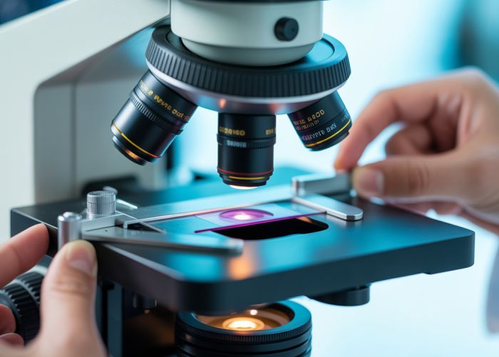
Microscopy, a technique essential in fields like histology, heavily relies on the effective slide microscope function. Carl Zeiss AG, a prominent manufacturer, contributes significantly to the advancement of these instruments. Understanding the various components involved in a slide microscope function, such as the objective lens, is crucial for accurate image acquisition and analysis.

Optimizing Article Layout for "Unlock the Slide Microscope Function: Guide & Tips!"
This document outlines the ideal article layout for a comprehensive guide on unlocking the "slide microscope function," focusing on clarity, user-friendliness, and search engine optimization (SEO).
1. Introduction: Grasping the Fundamentals
- Purpose: To engage the reader and clearly define the scope of the article.
- Content:
- Start with a relatable scenario (e.g., struggling to view slides clearly).
- Briefly explain what a slide microscope is and its common applications (e.g., education, research, diagnostics).
- Clearly define the "slide microscope function" – emphasize the core principles of proper slide preparation, focusing, and illumination for optimal viewing.
- Outline the article’s structure and the benefits readers will gain (e.g., improved slide viewing, better understanding of microscopic samples).
- Include a clear statement containing the primary keyword "slide microscope function" early in the introduction (e.g., "This guide will help you unlock the full potential of the slide microscope function.").
2. Preparing Your Microscope Slide: The Foundation for Success
- Purpose: To detail the necessary steps for creating high-quality microscope slides.
-
Content:
- Emphasize the importance of proper slide preparation for achieving a clear image.
- List necessary materials (e.g., slides, coverslips, staining solutions (if applicable), mounting medium).
- Provide step-by-step instructions for different preparation methods (e.g., wet mount, dry mount, stained slides).
2.1. Wet Mount Preparation
- Detailed instructions on preparing a wet mount.
- Diagram or image illustrating the process.
- Tips for preventing air bubbles.
2.2. Dry Mount Preparation
- Detailed instructions on preparing a dry mount.
- Image demonstrating the placement of the sample.
- Considerations for delicate specimens.
2.3. Staining Techniques (Optional)
- If applicable: A brief overview of common staining techniques.
- Mention specific stains relevant to common applications (e.g., Gram stain for bacteria).
- Link to more detailed resources on staining procedures.
2.4 Choosing the Right Mounting Medium
- Explain different types of mounting medium.
- Advise on selecting the best medium based on sample type.
3. Mastering the Microscope’s Focus: Clarity is Key
- Purpose: To guide the reader through the process of achieving a sharp, clear image.
-
Content:
- Explain the function of the coarse and fine focus knobs.
- Provide a step-by-step guide to focusing the microscope, starting with the lowest power objective.
- Address common focusing problems (e.g., difficulty finding the sample, blurry images).
3.1. Using the Coarse Adjustment Knob
- Explain how to use the coarse adjustment knob to initially bring the sample into focus.
- Emphasize the importance of careful movements to avoid damaging the objective or slide.
3.2. Fine-Tuning with the Fine Adjustment Knob
- Detail the use of the fine adjustment knob for achieving precise focus.
- Explain how to compensate for differences in refractive index.
3.3. Resolving Common Focusing Issues
Issue Possible Cause Solution Difficulty finding the sample Sample not properly centered Ensure the sample is centered on the slide and directly under the objective lens. Blurry Image Dirty objective lens Clean the objective lens with lens paper. Image drifting out of focus Mechanical instability Ensure the microscope is on a stable surface and that all components are properly tightened.
4. Illuminating Your Sample: Optimizing Light for Clear Viewing
- Purpose: To explain the role of illumination in microscopy and provide tips for optimizing light settings.
-
Content:
- Explain the function of the light source, condenser, and diaphragm.
- Describe different types of illumination (e.g., brightfield, darkfield, phase contrast).
- Provide guidance on adjusting the light settings for different types of samples and objectives.
4.1. Understanding the Light Source and Condenser
- Explain the types of light sources.
- Detail the role of the condenser in focusing the light.
4.2. Adjusting the Diaphragm for Optimal Contrast
- How to use the diaphragm to control the amount of light passing through the sample.
- Explain the relationship between diaphragm setting and image contrast.
4.3. Different Illumination Techniques: Brightfield, Darkfield (Briefly)
- Briefly describe Brightfield.
- Briefly describe Darkfield and Phase Contrast.
- Provide links for more information on each.
5. Troubleshooting Common Issues
- Purpose: To provide a resource for resolving common problems encountered while using a slide microscope.
-
Content:
- Present a table or list of common problems and their solutions.
- Address issues such as:
- Blurred images
- Dark images
- Air bubbles
- Drifting focus
- Uneven illumination
Problem Possible Cause(s) Solution(s) Blurred Image Dirty lenses, improper focusing, insufficient light Clean lenses with lens paper, refocus the microscope, adjust light settings. Dark Image Insufficient light, closed diaphragm Increase light intensity, open the diaphragm. Air Bubbles Improper slide preparation Prepare a new slide, ensuring to gently lower the coverslip at an angle. Drifting Focus Mechanical instability, temperature fluctuations Ensure the microscope is on a stable surface, allow the microscope to warm up before use. Uneven Illumination Misaligned light source, dirty condenser Align the light source, clean the condenser lens.
6. Maintaining Your Microscope: Ensuring Longevity
- Purpose: To provide guidance on the proper care and maintenance of a slide microscope.
-
Content:
- Explain the importance of regular cleaning and maintenance.
- Provide instructions on cleaning the lenses, stage, and other components.
- Address storage recommendations to prevent damage.
6.1. Cleaning the Lenses
- Step-by-step instructions on cleaning objective and eyepiece lenses.
- Emphasize the use of lens paper and appropriate cleaning solutions.
6.2. General Maintenance Tips
- Keeping the microscope covered when not in use.
- Avoiding exposure to dust and moisture.
So, hopefully, you found this guide helpful in understanding the slide microscope function. Go ahead and try out some of these tips, and let us know how it goes! Happy experimenting!