Understanding sinoatrial node function is paramount for comprehending cardiac physiology, a critical area of study for any aspiring cardiologist. Disruption of the sinoatrial node function often necessitates intervention, possibly including techniques advanced by the American Heart Association in arrhythmia management. Furthermore, accurate assessment of sinoatrial node function is achieved using diagnostic tools, like the electrocardiogram (ECG), to detect irregularities in heart rhythm. Impaired sinoatrial node function also highlights the importance of specialized centers, such as leading cardiac electrophysiology clinics, where innovative therapies restore proper heart rhythm.
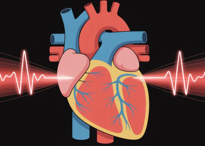
At the heart of our cardiovascular system lies a remarkable structure, often no larger than a grain of rice, yet possessing the extraordinary ability to orchestrate the rhythm of life itself. This is the sinoatrial node (SAN), the heart’s natural pacemaker.
It’s a specialized cluster of cells nestled within the right atrium, tirelessly working to ensure a consistent and coordinated heartbeat. Understanding the SAN is fundamental to grasping the intricacies of cardiac health and the profound impact of its proper function.
The Sinoatrial Node: Conductor of the Cardiac Orchestra
The primary function of the SAN is to initiate the electrical impulses that trigger each heartbeat. These impulses spread throughout the heart, causing the atria and ventricles to contract in a synchronized manner, efficiently pumping blood to the body.
Think of the SAN as the conductor of a complex orchestra. Without its precise timing and rhythmic cues, the entire system would fall into disarray.
The SAN generates electrical signals spontaneously, a process known as automaticity. This inherent ability to self-excite allows the heart to beat independently, even in the absence of external stimuli.
The rate at which these signals are generated determines our heart rate, which is constantly adjusted to meet the body’s changing demands.
The Ripple Effect: Impact on Overall Health
The proper functioning of the SAN is paramount to overall cardiovascular health. A healthy SAN ensures a stable and appropriate heart rate, providing the body with a consistent supply of oxygen and nutrients.
When the SAN malfunctions, the consequences can be significant. Irregular heart rhythms, known as arrhythmias, can lead to fatigue, dizziness, shortness of breath, and even life-threatening events.
A compromised SAN can disrupt the delicate balance of the cardiovascular system, affecting everything from blood pressure regulation to organ function. Recognizing the signs of SAN dysfunction and seeking timely medical attention is crucial for maintaining long-term well-being.
Anatomy and Location: Pinpointing the Heart’s Rhythmic Center
Now that we understand the SAN’s role as the heart’s conductor, it’s essential to delve into its physical attributes and precise location within the cardiac landscape. The SAN’s structure is intricately linked to its function, and appreciating its anatomical details provides a deeper understanding of its crucial role.
The Right Atrium: SAN’s Home
The sinoatrial node isn’t just floating around; it has a very specific address. It resides within the right atrium, one of the heart’s four chambers.
More specifically, it’s found near the junction where the superior vena cava – the large vein bringing deoxygenated blood from the upper body – enters the right atrium.
This strategic placement is vital for the efficient spread of electrical impulses throughout the heart.
Think of it as locating mission control right at the center of the operation; its central location allows for rapid signal transmission.
Pacemaker Cells: The SAN’s Microscopic Marvels
The SAN isn’t just a lump of ordinary heart tissue. It’s composed of specialized cells known as pacemaker cells or P-cells.
These cells are distinctly different from the contractile cardiomyocytes that make up the bulk of the heart muscle.
Pacemaker cells are smaller and contain fewer contractile elements.
Their unique characteristic lies in their ability to spontaneously generate electrical impulses, a property called automaticity.
This automaticity is due to specific ion channels within their cell membranes that allow for a slow, steady influx of ions, gradually building up to a threshold that triggers an action potential.
These action potentials then initiate the cascade of events leading to heart muscle contraction.
T-cells: Transitional Cells
The SAN also contains transitional cells, called T-cells, which are thought to facilitate the quick conduction of the impulse generated by P-cells, into the atrial muscle.
The SA Node Artery: Fueling the Rhythm
Like any active tissue, the SAN requires a dedicated blood supply to function optimally. This vital supply is provided by the SA node artery.
This artery, typically a branch of the right coronary artery, ensures that the SAN receives a constant flow of oxygen and nutrients.
Compromise to this artery leads to SAN disfunction.
In a significant portion of the population, the SA node artery originates from the right coronary artery, but variations exist, with some individuals having the artery originating from the left circumflex artery.
Regardless of its origin, the SA node artery’s patency is critical for maintaining the SAN’s function. Blockage or narrowing of this artery, due to conditions like atherosclerosis, can lead to SAN dysfunction and arrhythmias.
Physiology of SAN Function: How the Pacemaker Works
Having established the SAN’s physical location and cellular composition, we can now dissect the intricate mechanisms that allow it to function as the heart’s natural pacemaker. This involves understanding the generation of electrical impulses and their subsequent spread throughout the heart.
Action Potential Generation in Pacemaker Cells
The cornerstone of SAN function lies in its ability to spontaneously generate electrical impulses, known as action potentials. This unique property, called automaticity, distinguishes pacemaker cells from other cardiac cells. The process is driven by a complex interplay of ion channels within the cell membrane.
Role of Sodium, Calcium, and Potassium Channels
Ion channels are protein structures embedded in the cell membrane that act as selective gates, allowing specific ions to flow in or out of the cell. Sodium (Na+), calcium (Ca2+), and potassium (K+) channels play crucial roles in the generation of action potentials in pacemaker cells.
Unlike cardiomyocytes, pacemaker cells exhibit a slow, gradual depolarization during diastole (the resting phase between heartbeats). This is primarily due to the "funny current" (If), which is activated by hyperpolarization (a negative change in membrane potential).
The "Funny Current" (If) and Its Significance
The "funny current" (If) is a unique type of ion current that is carried mainly by sodium ions. It’s activated when the cell membrane becomes hyperpolarized, meaning more negative than its resting state.
This influx of sodium ions through If channels contributes to the slow, gradual depolarization of the pacemaker cell. This slow depolarization is what brings the cell closer to the threshold for firing an action potential.
Without the If current, the heart rate would be significantly slower and less responsive to changes in the body’s demands. The If current is considered a major player in heart rate regulation.
Depolarization and Repolarization
As the pacemaker cell slowly depolarizes, it eventually reaches a threshold potential. At this point, voltage-gated calcium channels open, allowing a rapid influx of calcium ions into the cell. This rapid influx of calcium causes the cell to rapidly depolarize.
This rapid influx of calcium causes the cell to rapidly depolarize, creating the upstroke of the action potential. Following depolarization, potassium channels open, allowing potassium ions to flow out of the cell. This outward flow of potassium ions causes the cell to repolarize, returning to its negative resting potential.
Hyperpolarization
After repolarization, the cell membrane potential becomes slightly more negative than its resting state, a state known as hyperpolarization. This hyperpolarization activates the "funny current" (If), initiating the cycle all over again.
Regulation of Heart Rate by the Autonomic Nervous System
While the SAN possesses inherent automaticity, its firing rate is finely tuned by the autonomic nervous system (ANS), which comprises the sympathetic and parasympathetic branches.
The Autonomic Nervous System and Its Branches
The autonomic nervous system (ANS) is a control system that acts largely unconsciously and regulates bodily functions such as the heart rate, digestion, respiratory rate, pupillary response, urination, and sexual arousal. It has two main branches: the sympathetic nervous system and the parasympathetic nervous system.
The Vagus Nerve
The parasympathetic nervous system, mediated by the vagus nerve, slows down heart rate. Vagal nerve stimulation releases acetylcholine, which acts on muscarinic receptors in the SAN to decrease the firing rate.
The Sympathetic Nervous System
Conversely, the sympathetic nervous system accelerates heart rate. Sympathetic nerve stimulation releases norepinephrine, which acts on beta-adrenergic receptors in the SAN to increase the firing rate.
Influence of Hormones
Hormones, such as epinephrine (adrenaline) released during stress or exercise, can also influence heart rate by acting on beta-adrenergic receptors in the SAN, similarly to norepinephrine.
Propagation of Signals in the Heart
Once the SAN generates an action potential, the electrical impulse must be efficiently propagated throughout the heart to trigger coordinated contraction.
Role of Internodal Pathways and Bachmann’s Bundle
The electrical signal spreads from the SAN through specialized pathways within the atria. These include the internodal pathways (anterior, middle, and posterior) and Bachmann’s bundle, which facilitates rapid conduction to the left atrium.
Interaction with Cardiomyocytes
As the electrical impulse spreads, it reaches the atrial cardiomyocytes, causing them to depolarize and contract. The impulse then travels to the atrioventricular (AV) node, where it is briefly delayed before being conducted to the ventricles via the bundle of His and Purkinje fibers.
Relationship Between Chronotropy, Inotropy, Cardiac Output, and Blood Pressure
The SAN’s control over heart rate (chronotropy) has a cascading effect on other crucial cardiovascular parameters. Increased heart rate, along with increased contractility (inotropy), leads to a higher cardiac output—the volume of blood pumped by the heart per minute.
Cardiac output, along with peripheral vascular resistance, determines blood pressure. Therefore, SAN dysfunction can significantly impact blood pressure regulation.
Role of the Cardiac Conduction System
The SAN is the heart’s primary pacemaker, but it relies on an intact cardiac conduction system to effectively distribute its electrical impulses throughout the heart. The AV node, bundle of His, and Purkinje fibers are all critical components of this system.
Disruptions in any part of the conduction system can lead to arrhythmias and compromise the heart’s ability to pump blood efficiently.
In summary, the SAN’s function is a masterpiece of biological engineering, involving intricate cellular mechanisms, hormonal influences, and coordinated electrical signaling. Understanding these physiological processes is crucial for appreciating the SAN’s vital role in maintaining cardiovascular health.
Measuring SAN Function: Diagnostic Tools for Assessment
Having explored the SAN’s intricate physiology and its dependence on a symphony of ion channels and regulatory influences, the question becomes: how do we assess whether this crucial pacemaker is functioning correctly? The evaluation of SAN function relies on a range of diagnostic tools, with the electrocardiogram (ECG/EKG) taking center stage.
The Electrocardiogram (ECG/EKG): A Window into SAN Activity
The electrocardiogram (ECG or EKG) serves as the primary non-invasive tool for assessing the sinoatrial node’s function. By recording the electrical activity of the heart from the body surface, the ECG provides valuable insights into the regularity, rate, and overall health of the heart’s rhythm.
Identifying P Waves: Atrial Depolarization Unveiled
The P wave on an ECG represents the electrical activity associated with atrial depolarization – the process by which the atria contract. Since the SAN is the primary initiator of this electrical activity, the presence, morphology, and timing of P waves are crucial indicators of SAN function.
A normal P wave suggests that the SAN is successfully initiating electrical impulses and that these impulses are being conducted through the atria in a coordinated manner.
Absent, inverted, or abnormally shaped P waves may indicate SAN dysfunction or ectopic atrial activity (electrical impulses originating from somewhere other than the SAN).
Analyzing RR Intervals: Unveiling Rhythmic Consistency
The RR interval represents the time between two consecutive R waves on the ECG, which correspond to ventricular contractions.
Analyzing the regularity and duration of RR intervals provides valuable information about the consistency of the heart’s rhythm.
In healthy individuals, the RR intervals are typically regular, reflecting the consistent firing of the SAN. Significant variations in RR intervals, known as arrhythmias, can suggest SAN dysfunction.
For example, a prolonged RR interval might indicate sinus bradycardia (a slow heart rate originating from the SAN), while an irregularly irregular RR interval could point to atrial fibrillation, where the SAN’s control is overridden by chaotic electrical activity in the atria.
Beyond the Standard ECG: Advanced Diagnostic Procedures
While the standard ECG provides a snapshot of heart rhythm, other diagnostic procedures offer more comprehensive and continuous assessments of SAN function.
Holter monitoring, for example, involves wearing a portable ECG recorder for 24-48 hours (or longer). This allows for the detection of intermittent arrhythmias or subtle SAN dysfunction that might be missed on a brief, standard ECG.
Electrophysiology studies (EPS) are invasive procedures that involve inserting catheters into the heart to directly record electrical activity and assess the function of the SAN and other components of the cardiac conduction system. EPS are typically reserved for complex cases or when more detailed information is needed to guide treatment decisions. These tests can pinpoint the exact location of conduction blocks or abnormal electrical pathways.
Analyzing the ECG allows clinicians to glean a significant amount of information regarding the heart’s electrical activity, but the story doesn’t end there. Aberrations in the P wave or inconsistencies in RR intervals can point to underlying issues within the sinoatrial node itself. When the SAN falters, a cascade of rhythm disturbances can arise, impacting the heart’s ability to efficiently pump blood. Let’s delve into these potential disorders and understand their ramifications.
Disorders of the Sinoatrial Node: Identifying Potential Problems
The sinoatrial node, though small, is indispensable. When it malfunctions, the consequences can range from subtle irregularities to life-threatening arrhythmias. Sinoatrial node dysfunction (also known as sick sinus syndrome), encompasses a spectrum of abnormalities in the heart’s rhythm, all stemming from a faulty pacemaker.
Understanding Sinoatrial Node Dysfunction
Sinoatrial node dysfunction isn’t a single disease, but rather a collection of conditions. These conditions all affect the SAN’s ability to generate and transmit electrical impulses effectively. The causes are varied, including age-related degeneration, underlying heart disease, certain medications, and, in some cases, genetic factors.
The manifestations of SAN dysfunction are diverse. Some individuals may experience periods of excessive slowness in their heart rate (bradycardia), while others might encounter alternating episodes of slow and fast heart rhythms (tachycardia-bradycardia syndrome). Still others might see the heart pause or skip beats entirely (sinus arrest or sinoatrial block).
Common Arrhythmias Associated with SAN Dysfunction
SAN dysfunction often manifests as various arrhythmias, which are irregular heartbeats. Understanding these arrhythmias is crucial for accurate diagnosis and effective management.
Sick Sinus Syndrome
As previously mentioned, sick sinus syndrome (SSS) is an overarching term. It describes several arrhythmias resulting from a malfunctioning SAN. These can include:
-
Sinus Bradycardia: An abnormally slow heart rate originating from the sinus node.
-
Sinus Arrest: The SAN temporarily stops firing, leading to pauses in the heart rhythm.
-
Sinoatrial Block: Electrical impulses generated by the SAN are unable to effectively reach the atria, resulting in skipped heartbeats.
-
Tachycardia-Bradycardia Syndrome: Alternating periods of rapid heart rates (such as atrial fibrillation or atrial flutter) and slow heart rates (bradycardia).
Bradycardia
Bradycardia, a heart rate slower than 60 beats per minute, can be a direct consequence of SAN dysfunction. While a slow heart rate can be normal in highly trained athletes, in other individuals, it may indicate a problem with the SAN’s ability to generate electrical impulses at an appropriate rate.
Tachycardia
While SAN dysfunction is often associated with slow heart rates, it can paradoxically also lead to rapid heart rates (tachycardia). This can occur in tachycardia-bradycardia syndrome. Here rapid atrial arrhythmias, like atrial fibrillation, are followed by abnormally slow rates.
Atrial Fibrillation
Atrial fibrillation (AFib) is a common arrhythmia characterized by rapid, irregular electrical activity in the atria. While AFib can arise independently of SAN dysfunction, the two can coexist. SAN dysfunction can sometimes predispose individuals to developing AFib.
Heart Block
Heart block, in the context of SAN dysfunction, typically refers to sinoatrial block. Here, electrical impulses from the SAN are unable to conduct properly to the atria. This results in missed beats and an irregular heart rhythm. More advanced heart blocks are typically located lower in the conduction system, such as the AV node or His-Purkinje system.
Impact on Cardiac Physiology
The effects of SAN dysfunction on cardiac physiology are significant. A consistently slow heart rate reduces cardiac output, leading to fatigue, dizziness, and shortness of breath. Erratic heart rhythms compromise the heart’s ability to fill and pump blood effectively. Over time, poorly managed SAN dysfunction can contribute to heart failure and increase the risk of stroke.
It is essential to promptly recognize and treat SAN dysfunction. Doing so helps restore a stable heart rhythm. Treatments can also alleviate symptoms and improve overall cardiovascular health.
Treatment Strategies for SAN Dysfunction: Restoring Rhythm and Function
Identifying the underlying cause and severity of sinoatrial node (SAN) dysfunction is only the first step. Crafting an effective treatment plan is crucial to alleviate symptoms, improve quality of life, and prevent potentially life-threatening complications. The approach often involves a multifaceted strategy, incorporating lifestyle adjustments, pharmacological interventions, and, in certain instances, pacemaker implantation.
Lifestyle Modifications: A Foundation for Heart Health
While lifestyle changes alone may not resolve SAN dysfunction entirely, they play a vital role in managing symptoms and optimizing overall cardiovascular well-being.
These modifications aim to reduce the burden on the heart and minimize factors that could exacerbate arrhythmias. Smoking cessation is paramount, as nicotine and other chemicals in cigarettes can damage the heart and blood vessels, further disrupting the SAN’s delicate electrical activity.
Moderating alcohol consumption is also advisable, as excessive alcohol intake can trigger atrial fibrillation and other arrhythmias.
Regular, moderate-intensity exercise, such as brisk walking or cycling, can improve cardiovascular fitness and help regulate heart rate.
It’s essential to consult with a physician before starting any new exercise program, particularly for individuals with pre-existing heart conditions.
Stress management techniques, like yoga, meditation, or deep breathing exercises, can help lower blood pressure and reduce the risk of arrhythmias associated with stress. A balanced diet, low in saturated and trans fats, cholesterol, and sodium, is also recommended to support overall heart health.
Medications: Managing Heart Rate and Arrhythmias
Pharmacological interventions aim to control heart rate, prevent blood clots (in the case of atrial fibrillation), and manage associated symptoms.
Medications are often used in conjunction with lifestyle modifications to achieve optimal results.
For individuals experiencing bradycardia (slow heart rate), medications may be adjusted or discontinued if they are contributing to the problem. In some cases, medications like atropine may be used temporarily to increase heart rate in emergency situations, but they are not a long-term solution for SAN dysfunction.
For those with tachycardia (fast heart rate) or atrial fibrillation, beta-blockers or calcium channel blockers can help slow the heart rate and control palpitations. Antiarrhythmic drugs, such as amiodarone or sotalol, may be prescribed to prevent or suppress atrial fibrillation, but these medications have potential side effects and require careful monitoring.
Anticoagulants, like warfarin or direct oral anticoagulants (DOACs), are often prescribed to individuals with atrial fibrillation to reduce the risk of stroke, a serious complication of this arrhythmia.
Pacemaker Implantation: Restoring Rhythmic Stability
In cases where lifestyle modifications and medications are insufficient to control symptoms or prevent life-threatening arrhythmias, pacemaker implantation may be necessary.
When is a Pacemaker Necessary?
Pacemaker implantation is typically considered when individuals experience:
- Symptomatic bradycardia (slow heart rate causing fatigue, dizziness, or fainting).
- Significant pauses in heart rhythm (sinus arrest or sinoatrial block).
- Tachycardia-bradycardia syndrome (alternating episodes of slow and fast heart rates).
- Syncope (fainting) due to SAN dysfunction.
The decision to implant a pacemaker is made on an individual basis, taking into account the patient’s symptoms, the severity of their arrhythmias, and their overall health status.
Types and Functions of Pacemakers
A pacemaker is a small, battery-powered device implanted under the skin, usually near the collarbone. It delivers electrical impulses to the heart to stimulate contraction when the heart’s natural pacemaker (the SAN) is not functioning properly.
-
Single-chamber pacemakers have one lead placed in either the right atrium or the right ventricle.
-
Dual-chamber pacemakers have leads placed in both the right atrium and the right ventricle, allowing for more coordinated heart contractions.
-
Rate-responsive pacemakers can adjust the heart rate based on the individual’s activity level, increasing the rate during exercise and decreasing it during rest.
-
Leadless pacemakers are a newer type of pacemaker that are smaller and do not require leads. They are implanted directly into the right ventricle.
Modern pacemakers are highly sophisticated devices that can be programmed to meet the individual’s specific needs. Regular follow-up appointments with a cardiologist are essential to ensure the pacemaker is functioning properly and to make any necessary adjustments. Pacemaker implantation can significantly improve the quality of life for individuals with SAN dysfunction, alleviating symptoms and reducing the risk of serious complications.
Frequently Asked Questions About Sinoatrial Node Function
These frequently asked questions provide clarification on some key aspects of sinoatrial node function and its role in regulating heart rhythm.
What exactly does the sinoatrial node do?
The sinoatrial node (SA node) is the heart’s natural pacemaker. Located in the right atrium, it generates electrical impulses that initiate each heartbeat. This process is fundamental to ensuring proper sinoatrial node function, allowing the heart to beat regularly.
How does the sinoatrial node control heart rate?
The SA node automatically depolarizes, creating an electrical signal that spreads throughout the heart. The rate of this depolarization determines the heart rate. Factors like hormones and the autonomic nervous system influence sinoatrial node function, speeding it up or slowing it down as needed.
What happens if the sinoatrial node malfunctions?
A malfunctioning SA node can lead to various heart rhythm problems, including bradycardia (slow heart rate), tachycardia (fast heart rate), or irregular heartbeats. These issues can disrupt normal heart function and may require medical intervention to restore proper sinoatrial node function.
Can lifestyle factors impact sinoatrial node function?
Yes, lifestyle factors can influence the SA node. Smoking, excessive alcohol consumption, and chronic stress can negatively impact sinoatrial node function and contribute to irregular heart rhythms. Maintaining a healthy lifestyle is crucial for overall heart health and optimal SA node performance.
So, that’s the scoop on sinoatrial node function! Hopefully, you now have a better grasp on how it all works. Keep your heart healthy, and remember to stay curious!