The anatomical study of organs, particularly through sheep heart dissection, is crucial for students engaging with biology classrooms. Sheep hearts themselves present a readily accessible model. They are similar to human hearts and can be obtained from slaughterhouses. Understanding the techniques involved in sheep heart dissection provides a solid foundation for comprehending cardiovascular systems and their functions. This guide will equip you with the knowledge and steps needed to master this essential skill.
Dissecting Sheep Hearts: The Ultimate Guide! – Article Layout Breakdown
This guide aims to provide a comprehensive and accessible layout plan for an article focused on "sheep heart dissection." The structure is designed to maximize clarity, learning, and engagement for readers with varying levels of experience.
Introduction: Setting the Stage for Sheep Heart Dissection
- Purpose: Briefly explain why sheep heart dissection is a valuable learning experience. This could involve highlighting its relevance to understanding human anatomy, cardiovascular health, and the scientific method.
- Relevance: Connect the sheep heart to the human heart. Mention similarities in structure and function, emphasizing that dissecting a sheep heart provides tangible insights into how our hearts work.
- Brief Overview: Outline what the article will cover. This acts as a roadmap for the reader, setting expectations and encouraging them to continue.
- Example Snippet: "Welcome to the ultimate guide to sheep heart dissection! This hands-on activity provides an incredible opportunity to explore the complexities of the circulatory system. By dissecting a sheep heart, which shares many similarities with a human heart, you’ll gain a deeper understanding of how this vital organ functions. In this guide, we’ll cover everything from preparing for the dissection to identifying key anatomical structures and understanding their roles."
Materials and Preparation: Getting Ready for the Dissection
-
List of Materials: Provide a clear and concise list of all necessary materials. This should include both the heart itself and the tools required for dissection.
- Sheep heart (preserved)
- Dissection tray
- Dissection kit (scalpel, forceps, probes, scissors)
- Gloves
- Apron or lab coat
- Paper towels
- Safety glasses
- Water source for rinsing
- Safety Precautions: Emphasize the importance of safety during the dissection.
- Wearing gloves and safety glasses is mandatory.
- Handle scalpels and scissors with extreme care.
- Wash hands thoroughly after the dissection.
- Proper disposal of biological waste is crucial.
-
Preparing the Heart: Explain how to properly rinse and position the sheep heart for dissection.
- Gently rinse the heart under cool water to remove excess preservative.
- Orient the heart to identify the anterior (front) and posterior (back) surfaces.
- Locate the apex (pointed end) and the base (where major blood vessels attach).
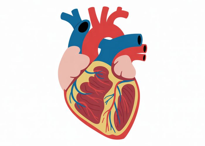
External Anatomy: Exploring the Outer Structures of the Sheep Heart
-
Identifying Major Blood Vessels: Describe how to identify and differentiate the major blood vessels connected to the heart. Use clear diagrams or photos.
- Aorta: The largest artery carrying oxygenated blood away from the heart.
- Pulmonary Artery: Carries deoxygenated blood to the lungs.
- Vena Cava (Superior and Inferior): Brings deoxygenated blood from the body back to the heart.
- Pulmonary Veins: Carry oxygenated blood from the lungs back to the heart.
-
Auricles: Explain the function of the auricles (ear-like flaps) located on the atria.
- These are small pouches that increase the capacity of the atria.
- Coronary Arteries and Veins: Point out the coronary arteries and veins, which supply blood to the heart muscle itself.
Internal Anatomy: Diving Deep Inside the Sheep Heart
-
Opening the Heart: Provide step-by-step instructions on how to carefully cut open the heart to reveal its internal structures.
- Make an incision along the anterior surface from the aorta to the apex.
- Continue the incision along the side of the heart, opening up the right ventricle.
- Repeat the process on the left side, cutting from the pulmonary artery to the apex.
-
Atria and Ventricles: Describe the differences between the atria (upper chambers) and the ventricles (lower chambers). Highlight the differences in wall thickness.
- Atria: Thinner walls; receive blood from veins.
- Ventricles: Thicker walls; pump blood to the lungs or the body. The left ventricle is significantly thicker than the right ventricle.
-
Valves: Explain the function of the heart valves and how to identify them.
- Tricuspid Valve (Right Atrioventricular Valve): Located between the right atrium and right ventricle.
- Mitral Valve (Bicuspid Valve/Left Atrioventricular Valve): Located between the left atrium and left ventricle.
- Pulmonary Valve: Located between the right ventricle and pulmonary artery.
- Aortic Valve: Located between the left ventricle and aorta.
-
Chordae Tendineae and Papillary Muscles: Describe the role of these structures in preventing the valves from inverting during ventricular contraction.
- Chordae Tendineae: Tendon-like cords that attach to the valve flaps.
- Papillary Muscles: Muscle projections that anchor the chordae tendineae.
Understanding Blood Flow: Tracing the Path Through the Heart
-
Step-by-Step Explanation: Provide a clear and concise explanation of the blood flow pathway through the heart. This section could benefit from visual aids, such as diagrams or flowcharts.
- Deoxygenated blood enters the right atrium via the vena cava.
- Blood flows through the tricuspid valve into the right ventricle.
- The right ventricle pumps blood through the pulmonary valve into the pulmonary artery.
- Blood travels to the lungs, where it becomes oxygenated.
- Oxygenated blood returns to the left atrium via the pulmonary veins.
- Blood flows through the mitral valve into the left ventricle.
- The left ventricle pumps blood through the aortic valve into the aorta.
- Blood is distributed throughout the body.
Troubleshooting and Common Errors: Addressing Potential Issues
- Identifying Structures: Difficulty in distinguishing between similar structures can be addressed through providing photographic comparisons.
- Cutting Errors: Guidance on avoiding damage to key structures when making incisions.
- Preservation Artifacts: Explanation of how preservation can alter the appearance of the heart and how to compensate.
Disposal and Cleanup: Concluding the Dissection
- Proper Disposal: Outline the correct procedures for disposing of the sheep heart and other biological waste.
- Cleaning the Workspace: Emphasize the importance of thoroughly cleaning and disinfecting the dissection tray and tools.
- Handwashing: Remind readers to wash their hands thoroughly with soap and water after the dissection.
FAQs: Sheep Heart Dissection Guide
Hopefully, our guide helped you perform a successful sheep heart dissection. Here are some frequently asked questions that might further clarify the process.
Why are sheep hearts used for dissection?
Sheep hearts are commonly used in dissection because they are structurally very similar to human hearts. Their size is also manageable for students, and they are readily available from biological supply companies at a reasonable cost. This makes a sheep heart dissection an accessible and effective way to learn about mammalian heart anatomy.
What’s the best way to identify the left and right ventricles?
The easiest way to distinguish the left and right ventricles is by examining their thickness. The left ventricle is much thicker because it has to pump blood to the entire body. The right ventricle only pumps blood to the lungs, so it is thinner. Also, feeling the internal septum will reveal a much stronger muscular wall on the left side of the sheep heart.
What are the functions of the chordae tendineae and papillary muscles?
The chordae tendineae ("heart strings") and papillary muscles work together to prevent the atrioventricular valves (mitral and tricuspid) from prolapsing or inverting back into the atria during ventricular contraction. They essentially anchor the valves, ensuring unidirectional blood flow. This is crucial during the sheep heart dissection process, as these internal structures should be easily visible.
What safety precautions should I take during a sheep heart dissection?
Always wear gloves and eye protection to prevent contact with preservatives and biological material. Use sharp dissection tools carefully and follow your instructor’s guidelines for proper disposal of the heart and materials. Wash your hands thoroughly with soap and water after completing the sheep heart dissection.
Alright, hopefully, you’re feeling confident enough to tackle that sheep heart dissection! It’s a fascinating process, and understanding the anatomy is something you can carry with you. Best of luck with your sheep heart dissection, and let me know if you have any questions!