The field of orthopedics recognizes sesamoid bones as integral components of biomechanical efficiency. Exploring human anatomy, specifically within the context of podiatry, helps define sesamoid bone by showcasing their crucial role in facilitating movement. The American Academy of Orthopaedic Surgeons provides extensive resources on conditions affecting these small but significant bones. Proper diagnosis, sometimes employing radiology techniques, is essential for addressing issues related to these bones.
Deep within the architecture of the human body lie numerous small, often overlooked, yet vitally important structures: the sesamoid bones.
These tiny bones, embedded within tendons, play a crucial role in biomechanics and skeletal function.
While present in various locations throughout the body, their significance is particularly pronounced in the foot, specifically at the base of the big toe.
These small structures are critical for activities ranging from walking to athletic endeavors.
However, these seemingly insignificant bones are prone to a variety of issues, from inflammation and chronic pain to fractures that can significantly impair mobility. Understanding their anatomy, function, and potential problems is paramount for anyone seeking to maintain optimal foot health.
What are Sesamoid Bones? Function and Purpose
Sesamoid bones are a unique type of bone, characterized by their encapsulation within a tendon.
Unlike most bones that connect directly at joints, sesamoids exist within the tendons of muscles.
This arrangement serves several essential purposes.
First, sesamoids act as pulleys, improving the angle of pull of the muscle and increasing its mechanical advantage.
Think of it as a rope passing over a wheel—the wheel (sesamoid) redirects the force, making the pull more efficient.
Second, they reduce friction between the tendon and underlying bone, preventing wear and tear.
Finally, sesamoid bones assist in weight-bearing and shock absorption, protecting the joint from excessive stress.
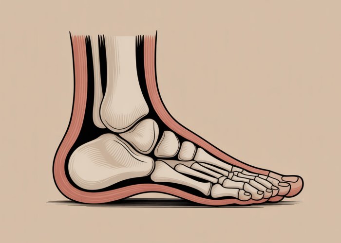
Location, Location, Location: Sesamoids in the Foot
While sesamoid bones exist in several locations throughout the body (hands, knees, etc.), they are perhaps most clinically relevant in the foot.
Specifically, two small sesamoid bones are located beneath the first metatarsophalangeal joint (MTPJ), which is the joint at the base of the big toe.
These two bones, the tibial and fibular sesamoids, are nestled within the flexor hallucis brevis tendon.
Their strategic positioning allows them to play a critical role in the biomechanics of the foot, particularly during activities like walking, running, and jumping.
Sesamoid Bone Problems: When Tiny Bones Cause Big Pain
Despite their small size, sesamoid bones are susceptible to a range of problems that can cause significant pain and disability.
Sesamoiditis, an inflammation of the sesamoids and surrounding tissues, is a common ailment, often resulting from overuse or repetitive stress.
Fractures, both acute and stress fractures, can also occur, typically caused by trauma or chronic overloading.
Additionally, dislocations and degenerative changes can affect these bones, leading to pain, limited mobility, and impaired function.
Recognizing the symptoms and risk factors associated with these conditions is vital for early diagnosis and treatment, ultimately promoting better outcomes and preventing chronic problems.
While sesamoid bones exist in several locations throughout the body (hands, knees, etc.), they are perhaps most clinically relevant in the foot. Their unique positioning and crucial biomechanical role in this weight-bearing structure make them particularly susceptible to injury and dysfunction. To fully appreciate their importance, a thorough understanding of their anatomical location is essential.
Anatomy 101: Where are the Sesamoid Bones Located?
The foot is a complex structure, housing a multitude of bones, ligaments, tendons, and muscles that work in harmony to provide support, propulsion, and balance. Within this intricate framework lie the sesamoid bones, two small, pea-shaped structures located on the plantar (bottom) aspect of the foot, directly beneath the head of the first metatarsal bone (the long bone leading to the big toe).
The Big Toe’s Tiny Guardians
More specifically, these sesamoids reside within the tendons of the flexor hallucis brevis muscle, which plays a vital role in flexing the big toe. One sesamoid is positioned on the medial (inner) side of the first metatarsal head, while the other is located on the lateral (outer) side.
These seemingly insignificant bones are cradled within grooves on the underside of the metatarsal head, ensuring their stability and proper alignment.
The Tendon-Bone-Joint Connection
The relationship between the sesamoid bones, the flexor hallucis brevis tendon, and the first metatarsophalangeal joint (MTP joint) is crucial to understanding their function. The sesamoids are essentially embedded within the tendon, acting as a fulcrum or a gliding surface for the tendon to slide over.
This arrangement significantly reduces friction and stress on the tendon as it moves during flexion and extension of the big toe. Furthermore, the sesamoids contribute to the overall stability of the first MTP joint, which is the joint connecting the metatarsal bone to the proximal phalanx (the first bone of the big toe).
The first MTP joint is critical for walking, running, and jumping. The sesamoid bones enhance the functionality of this joint.
Muscular Influences
Several other muscular structures surrounding the sesamoid bones and the first MTP joint also influence their function. The abductor hallucis muscle, located on the medial side of the foot, and the adductor hallucis muscle, located on the lateral side, both contribute to the movement and stability of the big toe.
These muscles, along with the flexor hallucis longus (which runs along the plantar aspect of the foot and inserts into the distal phalanx of the big toe), work in concert to control the complex motion of the first MTP joint. Any imbalance or dysfunction in these surrounding muscles can indirectly affect the sesamoid bones, potentially leading to pain and injury.
Understanding the intricate anatomy of the sesamoid bones and their surrounding structures is paramount for clinicians and individuals alike. Knowing the location of the sesamoids, their relationship to tendons and the MTP joint, and the influence of surrounding muscles provides a solid foundation for recognizing and addressing potential problems that may arise in this critical area of the foot.
The intricate anatomical arrangement sets the stage for understanding how these tiny bones contribute to the overall function and biomechanics of the foot. Their presence is not merely incidental; rather, they play a crucial role in optimizing movement, distributing weight, and ensuring the efficient operation of the lower limb.
Physiology and Biomechanics: How Sesamoid Bones Function
Sesamoid bones, though small, are mighty players in the foot’s biomechanical orchestra. Their function extends beyond simply occupying space; they are integral to reducing friction, improving muscle efficiency, and distributing weight during a variety of activities.
Reducing Friction and Enhancing Muscle Efficiency
One of the primary roles of sesamoid bones is to minimize friction between tendons and bones. By acting as a smooth gliding surface, they allow tendons, particularly the flexor hallucis brevis, to move more freely over bony prominences.
This reduction in friction translates directly into increased muscle efficiency. The muscle expends less energy overcoming resistance, allowing for smoother and more powerful movements.
Weight Distribution and Biomechanical Optimization
The sesamoid bones are strategically positioned to assist in weight distribution across the forefoot, specifically the first metatarsal head. During weight-bearing activities, such as walking, running, and jumping, the forces transmitted through the foot are considerable.
The sesamoids help to dissipate these forces, preventing excessive pressure on a single point.
This even distribution of weight is essential for maintaining balance, preventing injury, and optimizing biomechanics. The sesamoids act as a critical link in the kinetic chain, ensuring that forces are transferred efficiently throughout the foot and lower limb.
The Fulcrum Effect: Amplifying Muscle Power
Perhaps the most significant contribution of the sesamoid bones is their ability to act as a fulcrum for the flexor hallucis brevis tendon. By increasing the angle of pull of the tendon, the sesamoids effectively increase the force that the muscle can generate.
This fulcrum effect is analogous to using a wrench to tighten a bolt; the longer the handle, the greater the leverage and the more force that can be applied.
In the case of the foot, the sesamoid bones provide that extra leverage, allowing the flexor hallucis brevis to generate more force for flexing the big toe. This increased force is critical for push-off during walking and running, as well as for maintaining balance and stability.
The sesamoid bones of the foot, diligently performing their tasks of friction reduction and weight distribution, might seem like isolated architectural marvels. However, a closer look reveals a fascinating parallel with another crucial sesamoid bone in the human body: the patella, or kneecap.
The Patella Connection: A Comparative Look
The human body showcases ingenious design principles, often repeating effective solutions across different anatomical regions. One compelling example is the presence of sesamoid bones, small ossifications embedded within tendons, serving biomechanical functions. While the foot houses several sesamoids, a fascinating comparison can be drawn with the patella, or kneecap, the body’s largest sesamoid bone. Exploring this "patella connection" reveals shared principles of protection, biomechanical advantage, and overall physiological importance.
The Patella: The Body’s Largest Sesamoid Bone
It’s easy to overlook the significance of sesamoid bones due to their diminutive size. However, the patella immediately commands attention as the largest sesamoid bone in the human body. Situated within the quadriceps tendon, anterior to the knee joint, the patella’s size underscores its crucial role in lower limb biomechanics. Recognizing its classification as a sesamoid bone provides a valuable framework for understanding its functions and appreciating the underlying principles governing the action of these unique skeletal structures.
Parallel Functions: Protection and Biomechanical Advantage
While differing in size and specific anatomical location, the patella and the foot sesamoids share fundamental functional similarities. Both serve to protect underlying tendons and joints from stress and friction. The patella shields the knee joint and the quadriceps tendon, while the foot sesamoids safeguard the flexor hallucis brevis tendon and the first metatarsophalangeal joint.
Both sets of bones also provide a biomechanical advantage, optimizing joint movement and force transmission. The patella increases the leverage of the quadriceps muscle, allowing for more efficient knee extension. Similarly, the sesamoids in the foot enhance the power of the flexor hallucis brevis, facilitating push-off during gait. This shared principle of leveraging muscle force exemplifies the intelligent design inherent in the musculoskeletal system.
Emphasizing Common Physiological Function
The core physiological function uniting the patella and the foot sesamoids lies in their ability to improve joint mechanics and protect associated tendons. They exemplify nature’s approach to biomechanical engineering, demonstrating how relatively small structures can significantly enhance the function and resilience of larger joints and muscle groups. Both the patella and the sesamoids of the foot serve as fulcrums, changing the angle of the tendon’s pull and thereby increasing its mechanical advantage. This optimized biomechanics translates to greater efficiency, reduced stress on the joints, and improved overall performance of the lower limb.
Consider, for instance, the impact of a missing patella. The quadriceps muscle would lose a significant portion of its leverage, making activities like walking, running, and jumping considerably more difficult. Similarly, if the sesamoids in the foot were absent, the flexor hallucis brevis tendon would be more susceptible to injury, and the biomechanics of the first MTP joint would be compromised.
The comparative look between the patella and the foot sesamoids highlights a common theme of efficient force transmission and joint protection. This physiological kinship underscores the underlying principles of musculoskeletal design, revealing how strategically positioned sesamoid bones, regardless of size, contribute to the smooth, powerful, and resilient operation of the human body.
Hallux and First MTP Joint: The Core of Sesamoid Bone Action
As we’ve seen, sesamoid bones aren’t just tiny, incidental structures. Their influence is most profoundly felt at the big toe, or hallux, and the first metatarsophalangeal (MTP) joint. Understanding this relationship is crucial to appreciating their vital role in human movement.
The Hallux: A Foundation of Locomotion
The hallux, commonly known as the big toe, might seem like just another digit, but it’s a cornerstone of bipedal movement. It’s the final point of contact during push-off in each step, bearing a significant amount of body weight. This propulsive force relies heavily on the proper function of the first MTP joint.
A compromised hallux can disrupt gait, impacting everything from walking to running and jumping. The sesamoid bones play a critical role in ensuring the hallux can perform this function effectively.
Sesamoids: Facilitators of MTP Joint Function and Stability
The sesamoid bones are embedded within the tendons that cross the first MTP joint, serving as a crucial interface between the tendons and the joint itself. They’re positioned on the plantar aspect (the sole side) of the foot, directly beneath the head of the first metatarsal.
Their primary role is to enhance the smooth functioning and stability of the first MTP joint. They achieve this through several mechanisms:
-
Reducing Friction: Sesamoids act as a gliding surface, minimizing friction between the tendons and the bone. This allows for smooth, effortless movement during flexion and extension of the toe.
-
Increasing Mechanical Advantage: By acting as a fulcrum, sesamoids increase the leverage of the flexor hallucis brevis tendon. This enables greater force generation during push-off.
-
Weight Distribution: Sesamoids contribute to the distribution of weight across the first MTP joint, reducing stress on the metatarsal head.
Misalignment and Dysfunction: When Things Go Wrong
Given their critical role, any misalignment or dysfunction of the first MTP joint or the sesamoid bones can lead to a cascade of problems. This can manifest as:
-
Pain: Pain is a common symptom, often localized to the ball of the foot, specifically beneath the big toe joint.
-
Inflammation: Inflammation of the sesamoids (sesamoiditis) or surrounding tissues is a frequent occurrence.
-
Limited Range of Motion: Stiffness and reduced mobility in the first MTP joint can impede normal gait.
-
Hallux Valgus (Bunions): Although not directly caused by sesamoid dysfunction, bunions can alter the biomechanics of the first MTP joint, placing increased stress on the sesamoids.
-
Hallux Rigidus (Stiff Big Toe): Arthritis in the first MTP joint can limit motion, impacting the function of the sesamoids and causing pain.
Understanding the intricate relationship between the hallux, the first MTP joint, and the sesamoid bones is essential for diagnosing and addressing foot problems effectively. Recognizing the core role they play in locomotion allows for targeted interventions to restore function and alleviate pain.
Hallux and First MTP Joint: The Core of Sesamoid Bone Action
As we’ve seen, sesamoid bones aren’t just tiny, incidental structures. Their influence is most profoundly felt at the big toe, or hallux, and the first metatarsophalangeal (MTP) joint. Understanding this relationship is crucial to appreciating their vital role in human movement.
The Hallux: A Foundation of Locomotion
The hallux, commonly known as the big toe, might seem like just another digit, but it’s a cornerstone of bipedal movement. It’s the final point of contact during push-off in each step, bearing a significant amount of body weight. This propulsive force relies heavily on the proper function of the first MTP joint.
A compromised hallux can disrupt gait, impacting everything from walking to running and jumping. The sesamoid bones play a critical role in ensuring the hallux can perform this function effectively.
Sesamoids: Facilitators of MTP Joint Function and Stability
The sesamoid bones are embedded within the tendons that cross the first MTP joint, serving as a crucial interface between the tendons and the joint itself. They’re positioned on the plantar aspect (the sole side) of the foot, directly beneath the head of the first metatarsal.
Their primary role is to enhance the smooth functioning and stability of the first MTP joint. They achieve this through several mechanisms:
Reducing Friction: Sesamoids act as a gliding surface, minimizing friction between the tendons and the bone. This allows for smooth, effortless movement during flexion and extension of the toe.
Increasing Mechanical Advantage: By acting as a fulcrum, they increase the leverage of the tendons that bend the big toe. This makes push-off more efficient and powerful.
Weight Distribution: They help distribute weight evenly across the MTP joint, preventing excessive pressure on any one area. This is especially important during high-impact activities.
Shock Absorption: Sesamoids play a role in shock absorption, cushioning the joint from the impact of walking, running, and jumping.
Unfortunately, these small but mighty structures aren’t immune to problems. When subjected to undue stress or trauma, they can become inflamed, leading to a painful condition known as sesamoiditis.
Sesamoiditis: When These Tiny Bones Cause Big Pain
Sesamoiditis, simply put, is the inflammation of the sesamoid bones and the surrounding soft tissues. This condition predominantly affects the sesamoids located beneath the first metatarsal head in the foot, causing significant discomfort and hindering normal activity.
Understanding the causes, symptoms, and effective management strategies is paramount for anyone experiencing forefoot pain. Failing to address sesamoiditis can lead to chronic pain and limited mobility.
Defining Sesamoiditis: Inflammation and its Impact
Sesamoiditis is not merely a bone issue; it encompasses the inflammation of the entire functional unit—the bones, tendons, and bursae—around the first MTP joint. This inflammation disrupts the smooth gliding action of the tendons and compromises the joint’s shock-absorbing capabilities.
The pain associated with sesamoiditis arises from the constant irritation and pressure exerted on these inflamed structures with each step taken.
Symptoms: Recognizing the Signs of Trouble
The hallmark symptom of sesamoiditis is pain beneath the big toe joint. However, this pain often manifests in various ways:
- Gradual Onset: Unlike a fracture, sesamoiditis pain usually develops gradually over weeks or months.
- Aching Discomfort: The pain is often described as a dull ache that worsens with activity.
- Point Tenderness: Direct pressure on the sesamoid bones elicits sharp pain.
- Swelling and Stiffness: Mild swelling and stiffness may be present around the big toe joint.
- Difficulty Walking: Weight-bearing activities, especially those involving push-off, become painful and difficult.
It’s crucial to differentiate sesamoiditis from other conditions presenting similar symptoms. This requires careful clinical examination and potentially imaging studies to rule out fractures or turf toe.
Common Causes: Unraveling the Root of the Problem
Several factors can contribute to the development of sesamoiditis. Understanding these causes is crucial for prevention and effective management:
- Overuse: Repetitive stress from activities like running, dancing, and basketball can overload the sesamoid bones.
- High-Impact Activities: Sports involving frequent jumping and landing put excessive force on the forefoot.
- Improper Footwear: Shoes lacking adequate cushioning or support can increase stress on the sesamoids.
- Foot Structure: Individuals with high arches or prominent metatarsal heads may be predisposed to sesamoiditis.
- Sudden Increase in Activity: Ramping up training intensity too quickly without proper conditioning can overwhelm the sesamoids.
It is paramount to identify and address these contributing factors to facilitate healing and prevent recurrence. Modifying training regimens, choosing appropriate footwear, and addressing biomechanical imbalances are key steps in managing sesamoiditis effectively.
Hallux and First MTP Joint: The Core of Sesamoid Bone Action
As we’ve seen, sesamoid bones aren’t just tiny, incidental structures. Their influence is most profoundly felt at the big toe, or hallux, and the first metatarsophalangeal (MTP) joint. Understanding this relationship is crucial to appreciating their vital role in human movement.
The Hallux: A Foundation of Locomotion
The hallux, commonly known as the big toe, might seem like just another digit, but it’s a cornerstone of bipedal movement. It’s the final point of contact during push-off in each step, bearing a significant amount of body weight. This propulsive force relies heavily on the proper function of the first MTP joint.
A compromised hallux can disrupt gait, impacting everything from walking to running and jumping. The sesamoid bones play a critical role in ensuring the hallux can perform this function effectively.
Sesamoids: Facilitators of MTP Joint Function and Stability
The sesamoid bones are embedded within the tendons that cross the first MTP joint, serving as a crucial interface between the tendons and the joint itself. They’re positioned on the plantar aspect (the sole side) of the foot, directly beneath the head of the first metatarsal.
Their primary role is to enhance the smooth functioning and stability of the first MTP joint. They achieve this through several mechanisms:
Reducing Friction: Sesamoids act as a gliding surface, minimizing friction between the tendons and the bone. This allows for smooth, effortless movement during flexion and extension of the toe.
Increasing Mechanical Advantage: By acting as a fulcrum, sesamoids increase the leverage of the tendons that move the big toe. This is particularly important during push-off, allowing for a more powerful and efficient stride.
Now that we’ve established the integral role sesamoid bones play in the mechanics of the foot, particularly within the critical interplay of the hallux and the first MTP joint, it’s essential to consider the potential risks these small but mighty structures face. These bones, vital for movement and stability, are susceptible to injury, with fractures representing a significant concern.
Fractures and Stress Fractures: Recognizing the Risks
Sesamoid bone fractures represent a disruption in the integrity of these small but vital components of the foot. These fractures can manifest in different forms, each with its own distinct cause and presentation.
Differentiating between acute fractures and stress fractures is crucial for accurate diagnosis and effective treatment.
Acute vs. Stress Fractures: A Tale of Two Breaks
Sesamoid fractures generally fall into two categories: acute fractures and stress fractures. Understanding the distinction between them is vital for proper diagnosis and treatment.
Acute fractures are typically the result of a single, traumatic event. Think of a direct blow to the foot or a sudden, forceful hyperextension of the big toe.
Stress fractures, on the other hand, are insidious. They develop gradually over time due to repetitive stress and overuse.
Acute Sesamoid Fractures:
Acute fractures are often more straightforward to diagnose due to their association with a specific injury. They represent a sudden break in the bone, often accompanied by immediate and intense pain.
These fractures demand prompt medical attention.
Sesamoid Stress Fractures:
Stress fractures can be trickier to identify. The pain may start subtly and gradually worsen over weeks or months.
Athletes, especially runners and dancers, are particularly vulnerable to sesamoid stress fractures. They occur when the bones are subjected to repetitive, high-impact forces without adequate recovery time.
Etiology: Understanding the Root Causes
The cause of a sesamoid fracture dictates the nature of the break. Acute fractures are almost always trauma-induced, like a direct impact or forceful twisting.
Stress fractures, however, arise from a more complex interplay of factors.
- Repetitive stress: High-impact activities without adequate rest periods.
- Improper footwear: Shoes lacking sufficient cushioning or support.
- Biomechanical abnormalities: Foot structure or gait issues that place excessive stress on the sesamoids.
- Sudden increase in activity: Abruptly increasing the intensity or duration of training.
Recognizing the Signs: Symptoms of Sesamoid Fractures
The symptoms of a sesamoid fracture can vary depending on the type and severity of the fracture. It’s important to be aware of these signs to seek timely medical attention.
Common symptoms include:
- Pain: Localized pain in the ball of the foot, near the big toe joint. The pain may be sharp and intense with acute fractures or a dull ache that worsens with activity in stress fractures.
- Swelling: Inflammation and swelling around the sesamoid bones.
- Bruising: Discoloration of the skin due to bleeding under the surface, often seen with acute fractures.
- Difficulty weight-bearing: Pain and instability may make it difficult to put weight on the affected foot.
- Tenderness to touch: Pain upon palpation of the sesamoid bones.
- Stiffness: Limited range of motion in the big toe joint.
Diagnostic Tools: Unveiling the Fracture
Accurate diagnosis is paramount for effective management of sesamoid fractures. A thorough physical examination, combined with appropriate imaging techniques, is essential to confirm the diagnosis and rule out other potential causes of foot pain.
X-rays:
X-rays are typically the first-line imaging study for evaluating suspected sesamoid fractures. They can clearly visualize acute fractures and sometimes show evidence of stress fractures, although the latter may be more subtle.
It is crucial to get your foot checked out when you experience any of the listed symptoms to ensure the problem is addressed sooner than later.
Hallux and First MTP Joint: The Core of Sesamoid Bone Action
As we’ve seen, sesamoid bones aren’t just tiny, incidental structures. Their influence is most profoundly felt at the big toe, or hallux, and the first metatarsophalangeal (MTP) joint. Understanding this relationship is crucial to appreciating their vital role in human movement.
The Hallux: A Foundation of Locomotion
The hallux, commonly known as the big toe, might seem like just another digit, but it’s a cornerstone of bipedal movement. It’s the final point of contact during push-off in each step, bearing a significant amount of body weight. This propulsive force relies heavily on the proper function of the first MTP joint.
A compromised hallux can disrupt gait, impacting everything from walking to running and jumping. The sesamoid bones play a critical role in ensuring the hallux can perform this function effectively.
Sesamoids: Facilitators of MTP Joint Function and Stability
The sesamoid bones are embedded within the tendons that cross the first MTP joint, serving as a crucial interface between the tendons and the joint itself. They’re positioned on the plantar aspect (the sole side) of the foot, directly beneath the head of the first metatarsal.
Their primary role is to enhance the smooth functioning and stability of the first MTP joint. They achieve this through several mechanisms:
Reducing Friction: Sesamoids act as a gliding surface, minimizing friction between the tendons and the bone. This allows for smooth, effortless movement during flexion and extension of the toe.
Increasing Mechanical Advantage: By…
Diagnosis: Pinpointing the Problem
After understanding the intricate role sesamoid bones play in foot biomechanics, addressing any pain or discomfort in this area requires a precise diagnostic approach. Accurately identifying the source of the problem is paramount to effective treatment. This involves a multi-faceted strategy, combining a thorough physical examination with advanced imaging techniques, and employing differential diagnosis to rule out other potential conditions.
The Physical Examination: A Hands-On Assessment
The diagnostic journey typically begins with a comprehensive physical examination conducted by a qualified medical professional, such as a doctor or podiatrist. This initial assessment is crucial for understanding the patient’s symptoms and identifying potential problem areas.
During the examination, the doctor will carefully palpate the area around the first MTP joint, paying close attention to the location and characteristics of the pain. The goal is to pinpoint the exact source of the discomfort, whether it’s localized to one or both sesamoid bones, or if it radiates to surrounding tissues.
The doctor will also assess the range of motion of the big toe, noting any limitations or pain with movement. Observing the patient’s gait (walking pattern) can provide valuable insights into how the foot is functioning and whether the sesamoid bones are contributing to any biomechanical abnormalities.
Key Components of the Physical Exam
- Palpation: Gently feeling the area to identify tenderness, swelling, or masses.
- Range of Motion Assessment: Evaluating the flexibility and movement of the big toe joint.
- Gait Analysis: Observing the patient’s walking pattern for any abnormalities.
- Provocative Tests: Specific maneuvers designed to reproduce the patient’s pain and help confirm the diagnosis.
The Role of Imaging: Seeing Beneath the Surface
While the physical examination provides valuable clues, imaging techniques are often necessary to confirm the diagnosis and rule out other potential causes of foot pain. X-rays and Magnetic Resonance Imaging (MRI) are two commonly used imaging modalities that offer different perspectives on the sesamoid bones and surrounding structures.
X-Rays: Identifying Fractures and Dislocations
X-rays are a quick and readily available imaging technique that is particularly useful for identifying fractures and dislocations of the sesamoid bones. Because sesamoid bones are small, breaks can be subtle and difficult to detect.
X-rays can also help rule out other bony abnormalities, such as arthritis or bone spurs, that may be contributing to the patient’s symptoms.
MRI: A Deeper Dive into Soft Tissues
While X-rays excel at visualizing bone, MRI provides a more detailed view of the soft tissues surrounding the sesamoid bones, including tendons, ligaments, and cartilage. This is particularly valuable for detecting stress fractures, which may not be visible on X-rays until significant bone damage has occurred.
MRI can also identify soft tissue injuries, such as tendonitis (inflammation of the tendons) or ligament sprains, that may be contributing to the patient’s pain. In some cases, bone bruises (bone marrow edema) can be visualized with MRI after an injury.
Differential Diagnosis: Ruling Out Other Possibilities
Diagnosing sesamoid bone injuries requires a careful process of differential diagnosis, which involves considering other potential causes of foot pain and systematically ruling them out. Several conditions can mimic the symptoms of sesamoiditis or sesamoid fractures.
Common Conditions that Mimic Sesamoid Injuries:
- Turf Toe: A sprain of the ligaments around the big toe joint.
- Gout: A form of arthritis caused by a build-up of uric acid crystals in the joints.
- Morton’s Neuroma: A thickening of the tissue around a nerve in the foot.
- Plantar Fasciitis: Inflammation of the plantar fascia, a thick band of tissue that runs along the bottom of the foot.
- Capsulitis: Inflammation of the joint capsule.
By carefully considering these alternative diagnoses and using a combination of physical examination and imaging techniques, medical professionals can accurately pinpoint the source of the patient’s pain and develop an effective treatment plan. Accurate diagnosis is the cornerstone of successful management of sesamoid bone injuries.
Hallux and First MTP Joint: The Core of Sesamoid Bone Action
As we’ve seen, sesamoid bones aren’t just tiny, incidental structures. Their influence is most profoundly felt at the big toe, or hallux, and the first metatarsophalangeal (MTP) joint. Understanding this relationship is crucial to appreciating their vital role in human movement.
The Hallux: A Foundation of Locomotion
The hallux, commonly known as the big toe, might seem like just another digit, but it’s a cornerstone of bipedal movement. It’s the final point of contact during push-off in each step, bearing a significant amount of body weight. This propulsive force relies heavily on the proper function of the first MTP joint.
A compromised hallux can disrupt gait, impacting everything from walking to running and jumping. The sesamoid bones play a critical role in ensuring the hallux can perform this function effectively.
Sesamoids: Facilitators of MTP Joint Function and Stability
The sesamoid bones are embedded within the tendons that cross the first MTP joint, serving as a crucial interface between the tendons and the joint itself. They’re positioned on the plantar aspect (the sole side) of the foot, directly beneath the head of the first metatarsal.
Their primary role is to enhance the smooth functioning and stability of the first MTP joint. They achieve this through several mechanisms:
Reducing Friction: Sesamoids act as a gliding surface, minimizing friction between the tendons and the bone. This allows for smooth, effortless movement during flexion and extension of the toe.
Increasing Mechanical Advantage: By positioning themselves within the tendon, sesamoids increase the distance between the tendon and the joint’s axis of rotation. This increases the leverage and force that the muscle can exert.
The health and proper alignment of these small bones are therefore fundamental to overall foot function. When pain or injury arises, a spectrum of treatment options is available.
Treatment Options: From Conservative Care to Surgery
Addressing sesamoid bone injuries requires a nuanced approach. The optimal path hinges on the severity and nature of the injury, as well as the patient’s individual needs and activity level. Treatment strategies span the spectrum from conservative care to surgical intervention, each with its own set of considerations.
The Foundation of Healing: Conservative Treatment
Conservative treatment forms the cornerstone of managing most sesamoid injuries. The primary goals are to reduce pain and inflammation, protect the injured area, and promote healing. This multifaceted approach typically involves a combination of the following:
Rest, Ice, Compression, Elevation (RICE): The RICE protocol is often the first line of defense. Rest involves limiting weight-bearing activities to avoid further stress on the sesamoid bones.
Ice application helps reduce inflammation and pain. Compression, achieved with a bandage, provides support and minimizes swelling.
Elevation of the foot above heart level also aids in reducing swelling.
Pain Management: Over-the-counter pain relievers, such as ibuprofen or naproxen (NSAIDs), can effectively manage pain and inflammation. In some cases, a physician may prescribe stronger pain medications.
Immobilization: To further protect the injured sesamoid bones, immobilization may be necessary. This can be achieved with a walking boot, cast, or stiff-soled shoe. Immobilization reduces movement and allows the tissues to heal.
Finding Support: Orthotics and Insoles
Orthotics and insoles play a crucial role in supporting the foot and redistributing weight away from the injured sesamoid bones. They can be custom-made or over-the-counter, depending on the individual’s needs.
- Orthotics are custom-made shoe inserts designed to correct biomechanical imbalances in the foot. They can provide targeted support to the arch and offload pressure from the sesamoid bones.
- Insoles are over-the-counter shoe inserts that offer cushioning and support. They can help absorb shock and reduce stress on the foot.
By providing support and cushioning, orthotics and insoles can alleviate pain and promote healing.
When is Surgery Necessary?
Surgery is generally reserved for cases that do not respond to conservative treatment or when there is a severe fracture or dislocation. The decision to proceed with surgery is made after careful consideration of the patient’s symptoms, the severity of the injury, and their overall health.
- Severe Fractures: If a sesamoid bone is severely fractured or displaced, surgery may be necessary to stabilize the bone and restore proper alignment.
- Dislocations: In cases of sesamoid bone dislocation, surgery may be required to reposition the bone and stabilize the joint.
- Persistent Pain: When conservative treatments fail to provide relief from chronic pain, surgery may be considered as a last resort.
It is important to note that surgery carries inherent risks, and the recovery process can be lengthy. Therefore, it is essential to have a thorough discussion with a qualified orthopedic surgeon or podiatrist to determine if surgery is the appropriate treatment option. The goal is always to restore function and alleviate pain, but conservative methods should be exhausted first.
Conservative Treatment: RICE and Beyond
When sesamoid bones become a source of pain, the initial approach often involves a suite of conservative treatments. These non-surgical methods aim to reduce inflammation, alleviate pain, and promote natural healing processes. The acronym RICE (Rest, Ice, Compression, Elevation) forms the cornerstone of this approach, but effective management also extends to appropriate pain relief and immobilization when necessary.
The RICE Protocol: A Detailed Guide
RICE is a widely recognized and effective first-line treatment for numerous musculoskeletal injuries, including those affecting the sesamoid bones. Proper application of this protocol is critical for optimal outcomes.
Rest: Unloading the Affected Area
Rest is paramount in the early stages of sesamoiditis or fracture. It involves minimizing weight-bearing activities and avoiding movements that exacerbate pain. This might necessitate a temporary reduction in exercise intensity or a complete cessation of high-impact activities like running and jumping. The goal is to reduce stress on the sesamoid bones, allowing them to begin the healing process.
Ice: Cooling the Inflammation
Ice therapy is crucial for reducing inflammation and numbing pain. Apply an ice pack wrapped in a thin cloth (to protect the skin) to the affected area for 15-20 minutes at a time, several times a day. Consistency is key; regular ice applications, especially in the first few days following injury, can significantly diminish inflammation.
Compression: Supporting the Joint
Compression helps to minimize swelling and provide support to the injured area. An elastic bandage, applied snugly but not too tightly, can offer support and reduce fluid accumulation. Ensure the bandage doesn’t cut off circulation.
Elevation: Reducing Swelling
Elevating the foot above the heart promotes fluid drainage, further reducing swelling. This is particularly effective when combined with rest and ice. Prop the foot up on pillows while sitting or lying down to maximize the benefits of elevation.
Over-the-Counter Pain Relief: Managing Discomfort
Over-the-counter (OTC) pain relievers can play a valuable role in managing the pain associated with sesamoid bone injuries. Two common options are ibuprofen and acetaminophen.
Ibuprofen: An Anti-inflammatory Option
Ibuprofen is a nonsteroidal anti-inflammatory drug (NSAID) that helps to reduce both pain and inflammation. It’s important to follow the dosage instructions on the label and to be aware of potential side effects, such as stomach upset. Long-term use of NSAIDs should be discussed with a healthcare professional.
Acetaminophen: A Pain Reliever
Acetaminophen primarily relieves pain but has minimal anti-inflammatory effects. It’s a suitable option for individuals who cannot tolerate NSAIDs or when inflammation is not a primary concern. As with ibuprofen, adhere to recommended dosages to avoid potential liver damage.
Immobilization: Providing Stability and Protection
In some cases, immobilization may be necessary to further protect the sesamoid bones and promote healing. This can be achieved through the use of walking boots or casts.
Walking Boots: Allowing Limited Mobility
A walking boot provides support and stability while allowing for limited weight-bearing. It reduces stress on the sesamoid bones and helps to prevent further injury. The duration of walking boot use will vary depending on the severity of the injury.
Casts: Complete Immobilization
For more severe fractures or when complete immobilization is required, a cast may be necessary. A cast completely restricts movement of the foot and ankle, providing maximum protection and support. The decision to use a cast will be made by a healthcare professional based on the specific circumstances of the injury.
Inflammation Management: Reducing Pain and Swelling
Following the initial steps of RICE therapy, the next critical phase in managing sesamoid bone injuries revolves around effectively controlling inflammation. Prolonged inflammation not only exacerbates pain but also hinders the healing process, potentially leading to chronic issues. Therefore, a comprehensive approach targeting inflammation is crucial for achieving optimal outcomes.
The Role of Anti-Inflammatory Medications (NSAIDs)
Non-steroidal anti-inflammatory drugs (NSAIDs) are frequently employed to combat the inflammation associated with sesamoiditis and stress fractures. These medications, available both over-the-counter and by prescription, work by inhibiting the production of prostaglandins, substances that contribute to pain and inflammation.
Common examples include ibuprofen (Advil, Motrin) and naproxen (Aleve).
It’s important to note that while NSAIDs can provide significant relief, they are not without potential side effects. Prolonged use can increase the risk of gastrointestinal issues, such as ulcers and bleeding. Individuals with pre-existing kidney or heart conditions should exercise caution and consult their physician before starting NSAID therapy.
The optimal dosage and duration of treatment should be determined in consultation with a healthcare professional, who can assess individual risk factors and provide personalized recommendations. Remember, NSAIDs are a tool, not a cure, and should be used judiciously as part of a broader treatment strategy.
Topical Treatments: Targeted Relief
For individuals seeking a more localized approach to pain relief, topical treatments can offer a valuable alternative or adjunct to oral medications. These formulations, available as creams, gels, or patches, deliver medication directly to the affected area, minimizing systemic exposure and potential side effects.
Capsaicin cream, derived from chili peppers, works by depleting substance P, a neurotransmitter involved in pain signaling.
Topical NSAIDs, such as diclofenac gel, provide anti-inflammatory effects directly to the sesamoid bones and surrounding tissues.
Arnica is a homeopathic treatment believed to alleviate pain and swelling.
While topical treatments generally have a lower risk of systemic side effects compared to oral medications, it’s still essential to follow the product instructions carefully and discontinue use if any adverse reactions occur, such as skin irritation or allergic reactions.
Lifestyle Adjustments: Promoting Healing from Within
In addition to medications and topical treatments, several lifestyle adjustments can play a significant role in managing inflammation and promoting healing of sesamoid bone injuries.
Dietary Considerations
Adopting an anti-inflammatory diet can contribute to overall well-being and potentially reduce inflammation throughout the body. This involves emphasizing foods rich in omega-3 fatty acids (such as fatty fish, flaxseeds, and walnuts), fruits and vegetables (especially berries, leafy greens, and cruciferous vegetables), and spices like turmeric and ginger.
Conversely, limiting processed foods, sugary drinks, and unhealthy fats can help minimize inflammation.
Activity Modification
Adjusting activity levels is crucial to avoid exacerbating the injury and allow for proper healing. This might involve temporarily reducing the intensity or duration of weight-bearing activities, such as running or jumping. Cross-training activities that place less stress on the foot, such as swimming or cycling, can provide a way to maintain fitness without hindering the healing process.
Footwear and Orthotics
Wearing supportive footwear with adequate cushioning can help reduce stress on the sesamoid bones. Orthotics, custom or over-the-counter, can further improve foot biomechanics and provide additional support. A podiatrist can assess your foot structure and recommend appropriate footwear and orthotics.
Weight Management
Maintaining a healthy weight is essential for reducing the load on the sesamoid bones. Excess weight can contribute to increased stress and inflammation, delaying the healing process.
By incorporating these lifestyle adjustments into a comprehensive treatment plan, individuals can actively participate in their recovery and optimize their chances of a successful outcome. Remember, a holistic approach that addresses both the symptoms and the underlying causes of inflammation is key to long-term management of sesamoid bone injuries.
Topical treatments can offer considerable comfort, but they typically address the symptoms rather than the underlying cause of sesamoid bone issues. Likewise, over-the-counter medications and self-care measures may provide temporary relief. However, there comes a point when expert intervention becomes not just beneficial, but essential for proper diagnosis and effective treatment.
Orthopedics and Podiatry: Knowing When to Seek Expert Help
Navigating the world of foot and ankle pain can be challenging. Sesamoid bone injuries, in particular, often require specialized knowledge to diagnose and manage effectively. Knowing when to seek the expertise of an orthopedic surgeon or podiatrist is crucial for preventing long-term complications and ensuring a full recovery.
Understanding the Roles of Orthopedists and Podiatrists
Both orthopedic surgeons and podiatrists play vital roles in treating foot and ankle problems, but their training and focus differ slightly.
-
Orthopedic surgeons are medical doctors (MDs or DOs) specializing in the musculoskeletal system. They can diagnose and treat a wide range of conditions, including fractures, dislocations, arthritis, and sports injuries. They can perform surgery, prescribe medication, and recommend physical therapy.
-
Podiatrists, on the other hand, are doctors of podiatric medicine (DPMs) specializing in the foot and ankle. Their training focuses exclusively on the lower extremities. They are experts in diagnosing and treating foot and ankle conditions, including sesamoiditis, fractures, bunions, and hammertoes. They can also perform surgery, prescribe medication, and fit custom orthotics.
When Self-Treatment Isn’t Enough
Many sesamoid bone injuries can be managed effectively with conservative treatments, such as rest, ice, compression, and elevation (RICE therapy), along with over-the-counter pain relievers. However, certain situations warrant prompt consultation with a medical professional.
You should seek professional help if:
-
Your pain is severe or debilitating. If you are unable to bear weight on your foot or if the pain is interfering with your daily activities, it’s time to see a doctor.
-
Your symptoms persist or worsen despite home treatment. If you have been following the RICE protocol and taking pain medication for several days, but your symptoms are not improving, it’s essential to seek professional evaluation.
-
You suspect a fracture. If you have experienced a sudden injury to your foot, such as a fall or direct blow, and you are experiencing intense pain, swelling, and bruising, you may have a fracture. An X-ray is necessary to confirm the diagnosis.
-
You have numbness or tingling in your toes. This could indicate nerve damage, which requires prompt medical attention.
-
You have a history of foot problems or underlying medical conditions. Individuals with diabetes, peripheral neuropathy, or other conditions that affect the feet should seek professional care for any new foot pain or symptoms.
Recognizing the Need for Specialist Intervention
Certain circumstances necessitate consultation with a specialist, either an orthopedic surgeon or a podiatrist.
-
Diagnostic Uncertainty: If your primary care physician is unable to determine the cause of your foot pain, a specialist can provide a more thorough evaluation and utilize advanced imaging techniques, such as MRI, to reach an accurate diagnosis.
-
Complex or Severe Injuries: Severe fractures, dislocations, or soft tissue injuries may require surgical intervention or specialized treatment that is beyond the scope of general medical care.
-
Failed Conservative Treatment: If conservative treatments have failed to provide adequate relief, a specialist can explore other options, such as custom orthotics, injections, or surgery.
-
Chronic Pain: Persistent foot pain can significantly impact your quality of life. A specialist can help develop a comprehensive pain management plan that addresses the underlying cause of your pain and helps you regain function.
Proactive Foot Care: A Preventive Approach
While seeking expert help is crucial when problems arise, adopting a proactive approach to foot care can help prevent sesamoid bone injuries in the first place. This includes wearing supportive footwear, avoiding high-impact activities, and maintaining a healthy weight. Regular self-examination of your feet can also help you identify potential problems early on.
Ultimately, prioritizing foot health and recognizing the importance of timely professional intervention are key to maintaining an active and pain-free lifestyle. Don’t hesitate to seek the guidance of an orthopedic surgeon or podiatrist if you have concerns about your sesamoid bones or any other foot-related issues.
Topical treatments can offer considerable comfort, but they typically address the symptoms rather than the underlying cause of sesamoid bone issues. Likewise, over-the-counter medications and self-care measures may provide temporary relief. However, there comes a point when expert intervention becomes not just beneficial, but essential for proper diagnosis and effective treatment.
When conservative treatments fail to deliver lasting relief, and the pain associated with sesamoid bone issues becomes debilitating, the prospect of surgery may emerge. Understanding the various surgical options, the recovery journey, and potential risks is crucial for making an informed decision.
Surgery: A Last Resort
For many sesamoid bone problems, conservative care provides adequate relief. However, when pain persists despite diligent adherence to non-surgical treatments, or in cases of severe fractures or dislocations, surgical intervention may be considered. It’s important to view surgery as a last resort, carefully weighing the potential benefits against the inherent risks.
Surgical Procedures: Addressing Specific Issues
The type of surgery recommended will depend entirely on the nature and severity of the sesamoid bone problem. Here are a few common surgical approaches:
-
Sesamoidectomy: This involves the removal of one or both sesamoid bones. It is typically considered when conservative treatments have failed to alleviate pain caused by conditions like sesamoiditis or chronic sesamoid fractures. While it can provide relief, it also alters the biomechanics of the foot and can potentially lead to other problems.
-
Fracture Repair: If a sesamoid bone is fractured, surgical repair may be necessary, especially if the fracture is displaced or unstable. This typically involves using screws or pins to stabilize the bone fragments and promote healing.
-
Tendon Repair/Realignment: In some cases, sesamoid bone pain may be related to problems with the surrounding tendons, such as the flexor hallucis brevis. Surgery may be performed to repair or realign these tendons, which can help to restore normal sesamoid bone function.
-
Bone Grafting: In cases of nonunion fractures (where the bone fragments fail to heal), a bone graft may be used to stimulate bone growth and promote healing.
The best surgical approach is determined on a case-by-case basis, considering the patient’s overall health, activity level, and the specific characteristics of their sesamoid bone problem.
Navigating the Recovery Process
The recovery process following sesamoid bone surgery can be lengthy and demanding, requiring patience and adherence to the surgeon’s instructions. Here’s a general overview:
-
Weight-Bearing Restrictions: Following surgery, patients will typically be required to limit weight-bearing on the affected foot for several weeks or even months. This may involve using crutches, a walker, or a special boot.
-
Immobilization: The foot may be immobilized in a cast or brace to protect the healing bone and tissues. The duration of immobilization will vary depending on the type of surgery performed.
-
Physical Therapy: Once the initial healing phase is complete, physical therapy will be essential to restore range of motion, strength, and flexibility to the foot and ankle. A physical therapist will guide the patient through a series of exercises and stretches designed to improve function and reduce pain.
-
Gradual Return to Activity: Returning to normal activities should be a gradual process, guided by the surgeon and physical therapist. Rushing back too soon can increase the risk of re-injury or complications.
Understanding Potential Complications
As with any surgical procedure, there are potential complications associated with sesamoid bone surgery. While these complications are relatively rare, it’s crucial to be aware of them. Some potential complications include:
-
Infection: As with any surgery, there is a risk of infection at the surgical site. This can usually be treated with antibiotics.
-
Nerve Damage: There is a risk of damage to the nerves surrounding the sesamoid bones, which can lead to numbness, tingling, or pain.
-
Persistent Pain: In some cases, pain may persist even after surgery. This may be due to nerve damage, scar tissue formation, or other factors.
-
Hardware Problems: If screws or pins are used to stabilize a fracture, there is a risk of these devices loosening, breaking, or causing irritation.
-
Altered Biomechanics: Removal of a sesamoid bone (sesamoidectomy) can alter the biomechanics of the foot, potentially leading to pain or problems in other areas of the foot or ankle.
It is crucial to openly discuss the potential risks and complications with your surgeon before proceeding with surgery. A thorough understanding of these factors will help you make an informed decision about your treatment options.
Frequently Asked Questions About Sesamoid Bones
Sesamoid bones are small but important parts of your anatomy. Here are some common questions to help you understand them better.
Where are sesamoid bones typically found?
Sesamoid bones are usually located where tendons cross joints in the body. They’re embedded within those tendons. The most well-known example is the patella (kneecap), but they are also commonly found in the feet and hands.
What is the purpose of a sesamoid bone?
Sesamoid bones act like pulleys. They improve the efficiency of muscles by increasing their leverage. To define sesamoid bone simply, think of them as tiny protectors and enhancers of your movement.
What problems can arise with sesamoid bones?
Sesamoiditis, which is inflammation of the sesamoid bones and surrounding tissues, is a common issue. Fractures can also occur due to overuse or trauma. Pain and difficulty bearing weight are frequent symptoms.
Are sesamoid bones present in everyone?
While some sesamoid bones are consistently present (like the patella), the presence and location of others can vary. Some people may have more or fewer sesamoid bones, and they might be in slightly different locations. Knowing that not everyone has the same number and size of sesamoids is important.
So, that’s the scoop on sesamoid bones! Hopefully, you have a better understanding now, and you can now define sesamoid bone and are well informed about these little helpers.