The human eye, a remarkable biological instrument, owes its visual acuity to two primary photoreceptor cells: rod and cone. These cells, located in the retina, are responsible for converting light into electrical signals that the brain interprets as vision. Understanding the function of rod and cone not only enhances our appreciation for the complexities of sight but also informs advancements in fields like ophthalmology, aiding in the treatment of vision impairments.
Have you ever stopped to consider the sheer complexity of vision?
How does the simple act of opening your eyes translate into the rich, detailed world we perceive?
Vision is arguably our most relied-upon sense, shaping our understanding of the environment and influencing our interactions within it. From the mundane to the extraordinary, sight guides our steps, informs our decisions, and evokes a spectrum of emotions.
At the heart of this intricate process lie specialized cells within the retina, known as photoreceptors.
These microscopic structures, specifically rods and cones, are the gatekeepers of our visual experience.
They enable us to see and understand the world around us.
They convert light into signals that our brain interprets as images.
Without them, the world would be a blur, devoid of color and clarity.
The Primacy of Vision
Vision’s importance extends far beyond simply identifying objects.
It is integral to our spatial awareness, allowing us to navigate complex environments and interact with the world safely.
Our ability to recognize faces, read emotions, and appreciate art all hinges on the power of sight.
Consider the impact of impaired vision on daily life – the limitations it imposes on independence, the challenges it presents to learning and communication.
Vision is more than just a sense; it is a fundamental pillar of our existence.
Rods and Cones: The Dynamic Duo
The magic of sight hinges on the function of photoreceptors; the rods and cones.
These tiny but powerful cells orchestrate a complex process, transforming light into electrical signals that the brain can interpret.
Rods excel in low-light conditions, enabling us to see in the dimmest environments.
Cones, on the other hand, are responsible for our perception of color and fine detail in bright light.
These photoreceptors are not created equal; each plays a crucial role in providing a complete visual experience.
They do this by working together, constantly adapting to changing light conditions and providing the brain with a continuous stream of information.
Understanding the function of rods and cones is key to unlocking the secrets of sight.
The magic of sight hinges on the function of photoreceptors; the rods and cones. These tiny but powerful cells orchestrate a complex process, transforming light into electrical signals that our brain can interpret. But where exactly do these photoreceptors reside, and how are they organized within the eye to make vision possible?
The Eye’s Grand Design: Retina and Photoreceptor Overview
To understand how rods and cones function, we must first explore the architecture of the eye, with a particular focus on the retina. This delicate, multi-layered structure at the back of the eye is where the miracle of sight truly begins.
Unveiling the Retina’s Layers
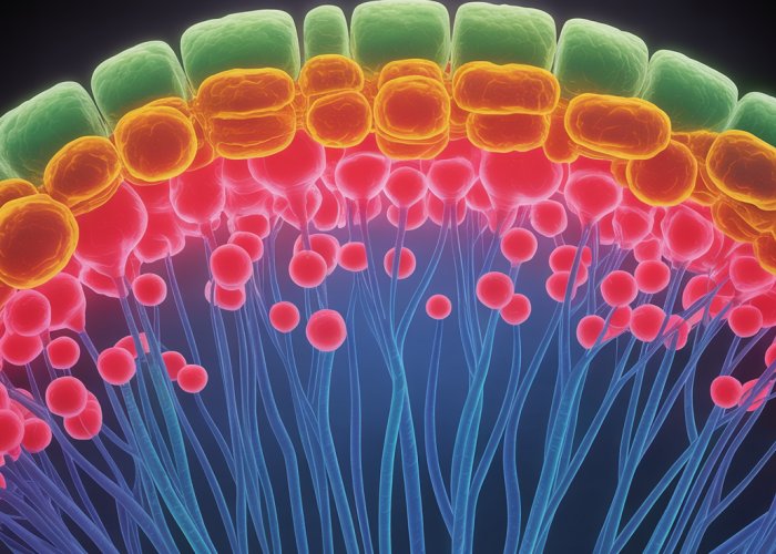
The retina isn’t a simple, uniform sheet of tissue; it’s a complex, layered structure, much like a sophisticated circuit board. Light must pass through several layers before reaching the photoreceptors, a seemingly counterintuitive design that highlights the intricate nature of the visual system.
From the inner limiting membrane (closest to the vitreous humor) to the outermost layer containing the photoreceptor cells, each layer plays a crucial role in processing visual information. These layers include:
-
Ganglion Cell Layer: Contains ganglion cells, whose axons form the optic nerve, transmitting signals to the brain.
-
Inner Plexiform Layer: Where ganglion cells synapse with bipolar and amacrine cells.
-
Inner Nuclear Layer: Contains the cell bodies of bipolar, amacrine, and horizontal cells.
-
Outer Plexiform Layer: Where photoreceptors synapse with bipolar and horizontal cells.
-
Outer Nuclear Layer: Contains the cell bodies of the photoreceptors (rods and cones).
-
Photoreceptor Layer: Contains the light-sensitive outer segments of rods and cones.
-
Retinal Pigment Epithelium (RPE): The outermost layer, providing support and nourishment to the photoreceptors. It also absorbs scattered light, preventing reflections that could blur vision.
This layered arrangement allows for complex processing of visual information before it even leaves the eye. The intermediate neurons, such as bipolar, horizontal, and amacrine cells, modulate the signals from the photoreceptors, enhancing contrast and adjusting to varying light levels.
Photoreceptors: The Gatekeepers of Light
Nestled within the photoreceptor layer of the retina are the stars of our visual show: rods and cones. These specialized cells are responsible for transducing light into electrical signals, the language of the nervous system. Without them, the rest of the eye’s sophisticated architecture would be for naught.
These cells are strategically positioned to capture incoming photons, initiating a cascade of biochemical events that ultimately lead to the generation of nerve impulses. These impulses then travel through the optic nerve to the visual cortex in the brain, where they are interpreted as images.
Rods vs. Cones: A Tale of Two Photoreceptors
While both rods and cones are photoreceptors, they are far from identical. They differ significantly in their location within the retina, their relative abundance, and their specific functions. Understanding these differences is crucial to appreciating the full scope of human vision.
Location, Location, Location
The distribution of rods and cones across the retina is far from uniform. Cones are highly concentrated in the fovea, the central part of the retina responsible for sharp, detailed vision. This explains why our visual acuity is highest when we look directly at an object.
Rods, on the other hand, are more prevalent in the periphery of the retina, outside the fovea. This distribution makes rods particularly important for peripheral vision and for detecting motion in our peripheral field of view.
A Numbers Game
In terms of sheer numbers, rods far outnumber cones in the human retina. There are approximately 120 million rods compared to only 6 million cones. This disparity reflects the importance of rods for night vision and peripheral awareness.
Functionality: Light, Color, and Clarity
The functional differences between rods and cones are perhaps the most significant. Rods are incredibly sensitive to light, allowing us to see in dim environments. They are responsible for scotopic vision, or night vision, but they do not detect color. This is why colors appear muted or absent in low-light conditions.
Cones, in contrast, require brighter light to function. They are responsible for photopic vision, or daylight vision, and, most importantly, for color perception. There are three types of cones, each sensitive to different wavelengths of light: red, green, and blue. It is the combined signals from these three types of cones that allow us to perceive the vast spectrum of colors that enrich our world.
In essence, rods and cones work together, each playing a distinct role in creating our visual experience. Rods provide us with sensitivity in low light and awareness of our surroundings, while cones give us the gift of color and clarity in brighter conditions. Understanding their individual contributions allows us to truly appreciate the grand design of the eye and the miracle of sight.
Unveiling the intricacies of the retina reveals a division of labor, a sophisticated specialization that allows us to navigate the world in varying degrees of illumination. While both rods and cones contribute to our visual experience, their roles are distinct. Now, let’s shift our focus to the unsung heroes of nighttime vision: the rods.
Rods: The Night Vision Specialists
In the realm of low-light environments, where colors fade and details blur, rods emerge as the dominant photoreceptors. They are responsible for our scotopic vision, or the ability to see in dim light. These highly sensitive cells are the key to navigating a starlit path or finding your way in a darkened room. They enable us to perceive the world even when light is scarce.
Rhodopsin: The Light-Sensitive Pigment
At the heart of a rod’s light-detecting prowess lies a remarkable molecule called rhodopsin, also known as visual purple.
Rhodopsin is a light-sensitive pigment that undergoes a chemical change when it absorbs even a single photon of light.
This change triggers a cascade of events that ultimately leads to an electrical signal being sent to the brain. The extraordinary sensitivity of rhodopsin allows rods to function in light intensities that would leave cones virtually dormant.
The Bleaching Process
When rhodopsin absorbs light, it undergoes a process called bleaching, where its structure changes and it loses its color. This bleached form is inactive and cannot respond to light.
To regain its light-sensitivity, rhodopsin must be regenerated through a process that requires time and specific enzymes. This regeneration process is crucial for dark adaptation.
Dark Adaptation: Adjusting to the Darkness
Have you ever experienced the initial blindness when stepping from a bright environment into a dark room? This is because dark adaptation is the process the eyes undergo to increase their sensitivity to low light levels.
Initially, the cones are overwhelmed by the sudden darkness, and the rods are still bleached from the previous bright light.
Over time, the cones become nonfunctional, and the rods gradually regenerate their rhodopsin. As more rhodopsin becomes available, the rods become increasingly sensitive, allowing us to see better in the dark.
This process can take several minutes, and full dark adaptation can take up to 30 minutes.
Peripheral Vision: A Rod-Dominated Domain
While cones are concentrated in the fovea, the central part of the retina responsible for sharp, detailed vision, rods are more abundant in the peripheral regions.
This distribution makes rods crucial for our peripheral vision, particularly in low-light conditions.
When navigating a dark environment, we often rely on our peripheral vision to detect movement or objects in our surroundings. This is because the rods in the periphery are more sensitive to dim light than the cones in the fovea.
Neural Interactions: Rods Communicating with the Brain
Rods don’t work in isolation.
They communicate with other neurons in the retina, such as bipolar cells and horizontal cells, to process visual information.
These interactions involve complex synaptic connections and the release of neurotransmitters.
The signals from rods are eventually transmitted to ganglion cells, whose axons form the optic nerve. This nerve carries the visual information to the brain for further processing and interpretation.
Rods have lower spatial resolution, meaning that many rods converge onto a single ganglion cell. While this increases light sensitivity, it comes at the expense of visual acuity.
Rods diligently illuminate our low-light world, but they paint it in shades of gray. For the vibrant hues and sharp details of daylight, we rely on a different set of photoreceptors.
Cones: Masters of Color and Clarity
While rods excel in the realm of darkness, cones reign supreme in bright light, bringing color and sharpness to our vision. They are the workhorses of photopic vision, our ability to see in well-lit environments. Cones enable us to appreciate the world in its full chromatic glory, from the fiery hues of a sunset to the delicate shades of a flower garden.
The Trio of Color: Red, Green, and Blue Cones
Unlike rods, which contain a single type of light-sensitive pigment, cones come in three distinct varieties, each tailored to respond to a specific range of wavelengths of light. These cone types are commonly referred to as red, green, and blue cones, although their sensitivity ranges are somewhat broader than these simple labels suggest.
-
Red cones are most sensitive to longer wavelengths, peaking in the reddish-yellow portion of the spectrum.
-
Green cones respond most strongly to medium wavelengths, corresponding to the green region of the spectrum.
-
Blue cones are most sensitive to shorter wavelengths, peaking in the blue-violet range.
It’s crucial to understand that these cones don’t exclusively detect red, green, or blue light. Instead, they exhibit varying degrees of sensitivity across a spectrum of wavelengths. The brain interprets the relative activity of these three cone types to perceive the full spectrum of color.
The Symphony of Color Vision
Color vision arises from the brain’s ability to process the signals sent by the three types of cones.
When light enters the eye, it stimulates the red, green, and blue cones to varying degrees, depending on its spectral composition. For instance, yellow light stimulates both red and green cones, but not blue cones.
The brain then interprets this pattern of stimulation to perceive the color yellow.
This process of mixing signals from different cone types allows us to perceive a vast array of colors, far beyond just red, green, and blue. It’s a remarkable feat of neural processing that enables us to appreciate the richness and complexity of the visual world.
Visual Acuity and the Fovea’s Central Role
The fovea, a small pit located in the center of the macula (the central area of the retina), is the region of the retina responsible for our sharpest, most detailed vision. What sets the fovea apart is its extremely high concentration of cones, with virtually no rods present.
This dense packing of cones allows for maximum spatial resolution, meaning we can discern fine details with greater clarity. Each cone in the fovea connects to its own ganglion cell, enabling the brain to receive a highly detailed representation of the central visual field.
The high concentration of cones in the fovea is the primary reason why our visual acuity is highest when we look directly at an object. It’s why we turn our heads to focus our gaze on what we want to see clearly.
Cone Communication: A Neural Relay Race
Like rods, cones don’t directly transmit information to the brain. Instead, they synapse with intermediate neurons, such as bipolar cells and horizontal cells, which further process and refine the visual signal.
Bipolar cells receive input from cones and transmit it to ganglion cells, the neurons whose axons form the optic nerve. Horizontal cells mediate lateral interactions between photoreceptors and bipolar cells, contributing to contrast enhancement and edge detection.
The signals from these intermediate neurons converge on ganglion cells, which act as the final relay station in the retina. The axons of ganglion cells bundle together to form the optic nerve, which carries the visual information to the brain for further processing. The intricate interplay between cones and these various neural cells ensures that the brain receives a comprehensive and refined representation of the visual scene, enabling us to perceive the world with remarkable clarity and color.
From Light to Sight: The Visual Pathway
The dance of light and photoreceptors within the retina is only the beginning of our visual experience. The information gathered by rods and cones must be translated, processed, and transmitted to the brain for interpretation. This intricate process involves a complex series of neural events, a journey from light’s initial impact to the conscious perception of sight.
Transduction: Converting Light into Electrical Signals
Rods and cones don’t simply "see" light; they transduce it, converting photons into electrical signals that the nervous system can understand.
This process begins with photoisomerization. Light triggers a change in the shape of retinal, a molecule bound to proteins within the photoreceptors.
This shape change initiates a cascade of biochemical events that ultimately lead to the closing of ion channels in the cell membrane.
The closing of these channels alters the electrical potential of the photoreceptor, creating a graded electrical signal.
The strength of this signal is proportional to the intensity of the light.
Intermediate Neurons: Refining the Visual Message
The signals generated by rods and cones don’t travel directly to the brain. Instead, they pass through a network of intermediate neurons within the retina, where the initial visual information is processed and refined.
Bipolar Cells: These cells receive input from photoreceptors and transmit it to ganglion cells. They act as crucial intermediaries, integrating and relaying information about light and dark.
Ganglion Cells: These are the final output neurons of the retina. Their axons converge to form the optic nerve. Ganglion cells receive input from bipolar cells and other retinal neurons. They then fire action potentials that transmit visual information to the brain. Different types of ganglion cells are specialized to detect different aspects of the visual scene, such as edges, motion, or color.
The Optic Nerve: A Highway to the Brain
The axons of all the ganglion cells converge at the optic disc, forming the optic nerve.
This nerve exits the eye and carries the encoded visual information towards the brain.
It’s a crucial link, a dense bundle of neural fibers transmitting the raw data that will ultimately be interpreted as sight.
At the optic chiasm, the optic nerves from each eye partially cross over, ensuring that each hemisphere of the brain receives information from both visual fields.
After the optic chiasm, the optic fibers continue as the optic tracts, heading towards the thalamus, a key relay station in the brain.
The Visual Cortex: Interpreting the World
From the thalamus, visual information is relayed to the visual cortex, located in the occipital lobe at the back of the brain.
This is where the magic of sight truly happens.
The visual cortex is organized hierarchically, with different areas responsible for processing different aspects of visual information, such as shape, color, motion, and depth.
Neurons in the visual cortex respond to specific features of the visual scene. Some neurons are activated by vertical lines, others by horizontal edges, and still others by particular colors or movements.
Through the intricate processing of the visual cortex, the electrical signals originating in the retina are transformed into the rich and meaningful visual world we experience.
It is through the remarkable journey along the visual pathway that mere photons are ultimately translated into perception, cognition, and awareness.
From the intricate pathways that translate light into understanding, we gain a new appreciation for the complexity of sight. But what happens when this delicate system is disrupted?
When Vision Falters: Understanding Impairments
The human visual system, a marvel of biological engineering, is unfortunately susceptible to a range of impairments. These conditions can arise from genetic factors, environmental influences, or the natural processes of aging, all impacting the function of rods, cones, and the neural pathways that support vision.
Achromatopsia: A World Without Color
Achromatopsia, often referred to as total color blindness, is a rare genetic condition characterized by a complete or near-complete absence of color vision. Individuals with achromatopsia typically possess non-functional or absent cones.
This results in a visual experience dominated by shades of gray.
Beyond color perception, achromatopsia often presents with reduced visual acuity, extreme light sensitivity (photophobia), and involuntary eye movements (nystagmus). The world appears blurry, washed out, and painfully bright.
Common Eye Diseases Affecting Photoreceptors
Several prevalent eye diseases specifically target and damage photoreceptor cells, leading to significant vision loss.
Macular Degeneration: Eroding Central Vision
Macular degeneration (MD) is a progressive eye disease that affects the macula, the central portion of the retina responsible for sharp, detailed vision.
Age-related macular degeneration (AMD), the most common form, primarily affects individuals over the age of 60. The deterioration of the macula leads to blurred or distorted central vision, making it difficult to read, drive, or recognize faces.
There are two main types of AMD:
-
Dry AMD: Characterized by the gradual thinning of the macula and the presence of drusen (small, yellow deposits) beneath the retina.
-
Wet AMD: Involves the abnormal growth of blood vessels beneath the retina, which can leak fluid and blood, causing rapid vision loss.
Retinitis Pigmentosa: A Slow March to Darkness
Retinitis pigmentosa (RP) is a group of inherited eye diseases that cause progressive degeneration of the retina, primarily affecting rod cells.
This leads to a gradual decline in night vision and peripheral vision, eventually resulting in tunnel vision.
Over time, cone cells can also be affected, leading to a decrease in color vision and visual acuity.
The progression of RP varies significantly among individuals, but it often leads to severe visual impairment or blindness.
The Impact of Neuronal Malfunction on Vision
Beyond diseases directly targeting photoreceptors, malfunctions in other neurons within the visual pathway can also significantly impair vision.
Damage to ganglion cells, for instance, can disrupt the transmission of visual information from the retina to the brain.
Furthermore, abnormalities in the visual cortex, the area of the brain responsible for processing visual information, can lead to a variety of visual disturbances.
These can range from difficulties in recognizing objects to the perception of distorted or absent images.
From the intricate pathways that translate light into understanding, we gain a new appreciation for the complexity of sight. But what happens when this delicate system is disrupted? We’ve touched on impairments like achromatopsia and macular degeneration, but now, let’s shift our focus to a critical nutrient essential for maintaining healthy vision: vitamin A.
Nourishing Sight: The Role of Vitamin A
Vitamin A, often hailed for its diverse health benefits, plays a particularly vital role in maintaining optimal vision. It acts as a key ingredient for the visual cycle. Without adequate vitamin A, the intricate dance of light and photoreceptors can falter, leading to impaired sight, especially under low-light conditions.
Vitamin A and Rhodopsin: An Inseparable Partnership
At the heart of rod cells lies rhodopsin, a light-sensitive pigment crucial for scotopic vision (night vision). Rhodopsin is not a single molecule; it’s a complex formed by combining a protein called opsin with retinal, a specific form of vitamin A.
When light strikes rhodopsin, retinal changes its shape, triggering a cascade of events that ultimately generate an electrical signal that is sent to the brain.
This transformation is essential for seeing in the dark. This entire process relies entirely on the presence of sufficient retinal derived from Vitamin A.
The Impact of Vitamin A Deficiency on Vision
A deficiency in vitamin A can have profound consequences for visual health, most notably affecting the function of rod cells and, consequently, night vision.
Night Blindness (Nyctalopia)
One of the earliest and most characteristic signs of vitamin A deficiency is nyctalopia, commonly known as night blindness.
This condition manifests as difficulty seeing in dim light or at night. Individuals with nyctalopia may struggle to navigate dimly lit environments, drive at night, or adjust to changes in light levels.
The lack of retinal hinders the regeneration of rhodopsin, leaving rod cells unable to effectively respond to low levels of light.
Xerophthalmia: A Cascade of Complications
Prolonged and severe vitamin A deficiency can lead to a condition called xerophthalmia. This is characterized by a range of ocular issues stemming from dryness of the conjunctiva and cornea.
Initially, xerophthalmia presents as conjunctival dryness (xerosis) and can progress to the formation of Bitot’s spots (foamy plaques on the conjunctiva).
If left untreated, xerophthalmia can ultimately lead to corneal ulceration, scarring, and irreversible blindness. This condition highlights the critical importance of vitamin A for maintaining the health and integrity of the eye’s surface.
Other Visual Consequences
Beyond night blindness and xerophthalmia, vitamin A deficiency can also contribute to other visual problems, including impaired color vision and reduced visual acuity.
While cones are less dependent on rhodopsin than rods, they still require vitamin A for optimal function.
Additionally, vitamin A plays a role in the overall health and maintenance of the retina, further emphasizing its importance for overall visual function.
In conclusion, vitamin A is an indispensable nutrient for maintaining healthy vision. Its role in rhodopsin production is critical for night vision, and its broader effects on the health of the cornea and retina underscore its importance for overall visual function. Addressing vitamin A deficiency is crucial for preventing and treating vision impairments.
FAQs About Rods and Cones
Hopefully, this sheds some light on rods and cones and how they work together to help us see. If you have more questions, feel free to ask!
What’s the main difference between rods and cones?
Rods primarily handle vision in low light conditions, enabling us to see in the dark. Cones are responsible for color vision and work best in bright light, allowing us to perceive detail.
Where are rods and cones located in the eye?
Rods and cones are photoreceptor cells located in the retina, which is the light-sensitive tissue at the back of your eye. They are not evenly distributed; cones are more concentrated in the fovea (the central part of the retina), while rods are more prevalent in the periphery.
How do rods and cones contribute to our perception of colors?
Cones are the key players in color vision. We have three types of cones, each sensitive to different wavelengths of light: red, green, and blue. Our brain interprets the signals from these cones to perceive the full spectrum of colors. Rods, on the other hand, do not contribute to color vision.
What happens if my rods or cones are damaged?
Damage to rods or cones can lead to various vision problems. For example, rod dysfunction can cause night blindness, making it difficult to see in dim light. Cone damage can lead to color blindness or decreased visual acuity in bright light, impacting your ability to see fine details.
So, there you have it – a glimpse into the fascinating world of rods and cones! Hopefully, you now appreciate these little guys a bit more. Go forth and see the world, and maybe even think about your rod and cone while you’re at it. Until next time!