The proteins primary structure, a fundamental aspect of molecular biology, dictates protein function through its specific amino acid sequence. The amino acid sequence, which defines the proteins primary structure, is ultimately determined by information encoded within DNA. Understanding the nuances of Edman degradation, a technique used to determine this sequence, allows scientists to map the blueprint of life’s building blocks. Moreover, the field of bioinformatics provides computational tools to analyze and predict proteins primary structure, further enhancing our knowledge of protein behavior. The proteins primary structure, therefore, serves as the foundation for the intricate three-dimensional architecture and functional capabilities studied in structural biology.
Proteins are the workhorses of the biological world, orchestrating virtually every process within living organisms. From catalyzing biochemical reactions to transporting molecules, providing structural support, and defending against pathogens, their functions are as diverse as life itself. Understanding these intricate roles begins with deciphering their fundamental building block: the primary structure.
This article delves into the significance of protein primary structure, the linear sequence of amino acids that dictates a protein’s unique identity and function. We will explore how this seemingly simple arrangement serves as the foundation for all higher-order structures and ultimately governs protein behavior.
The Ubiquitous Role of Proteins
Proteins are indispensable macromolecules. They participate in virtually every cellular process. Enzymes, antibodies, hormones, and structural components like collagen are all proteins, highlighting their multifaceted importance.
Their synthesis is meticulously controlled by the genetic code. It ensures that each protein is built with the precise sequence of amino acids required for its specific task. Errors in this sequence can have profound consequences, leading to disease or dysfunction.
The Primary Structure: A Foundation for Function
The primary structure of a protein is its amino acid sequence, linked together by peptide bonds. This sequence is not merely a random assortment. It’s a precisely ordered chain that dictates how the protein will fold, interact with other molecules, and ultimately perform its biological role.
Think of it as the blueprint for a complex machine. The sequence determines the three-dimensional shape of the protein. This then dictates its interactions with other molecules. Even a single amino acid change can disrupt the protein’s structure and function.
Implications for Medicine and Biotechnology
The study of protein primary structure is crucial for advancing medicine and biotechnology. Understanding the sequence of disease-related proteins allows us to:
- Develop targeted therapies.
- Design novel proteins with enhanced functions.
- Unravel the molecular mechanisms of disease.
For example, identifying mutations in proteins that cause cancer can lead to the development of drugs that specifically target those mutated proteins.
Furthermore, knowledge of protein sequences enables the creation of synthetic proteins and peptides for therapeutic or industrial applications. Understanding protein primary structure is therefore not just an academic pursuit. It is a powerful tool with real-world implications for improving human health and advancing technological innovation.
Think of it as the blueprint for a complex machine. The sequence determines the three-dimensional shape of the protein. This then dictates its interactions with other molecules. Even a single amino acid change can have dramatic consequences. But what exactly are these amino acids, and how do they contribute to the astonishing diversity and functionality of proteins?
The Building Blocks: Amino Acids and Their Diversity
Proteins, the molecular machines of life, are constructed from a set of 20 standard amino acids. These amino acids are the fundamental units that, when linked together in a specific sequence, determine a protein’s unique characteristics and biological activity. Understanding their structure and properties is crucial to grasping how proteins function.
The General Structure of Amino Acids
Each amino acid shares a common core structure.
It consists of a central carbon atom (the α-carbon) bonded to four different groups:
- An amino group (-NH2).
- A carboxyl group (-COOH).
- A hydrogen atom (-H).
- And a distinctive side chain (also known as an R-group).
It is the side chain that differentiates each of the 20 standard amino acids.
This seemingly small variation is the source of immense chemical diversity.
The Diversity of Side Chains
The side chains of amino acids vary in size, shape, charge, hydrogen-bonding capacity, hydrophobicity, and chemical reactivity.
This variability dictates how each amino acid interacts with its environment and with other amino acids within the protein.
The unique properties of each side chain play a critical role in determining a protein’s three-dimensional structure.
It also determines its interactions with other molecules.
Categories of Amino Acids and Their Properties
Amino acids are often categorized based on the properties of their side chains. The main categories are:
-
Nonpolar (Hydrophobic) Amino Acids: These amino acids have side chains that are primarily composed of hydrocarbons. They tend to cluster together in the interior of a protein, away from water, through hydrophobic interactions. Examples include alanine, valine, leucine, isoleucine, phenylalanine, tryptophan, and methionine.
-
Polar (Hydrophilic) Amino Acids: These amino acids have side chains that contain atoms (like oxygen or nitrogen) that create a partial charge, allowing them to form hydrogen bonds with water. They are typically found on the surface of a protein. Examples include serine, threonine, cysteine, tyrosine, asparagine, and glutamine.
-
Acidic (Negatively Charged) Amino Acids: These amino acids have side chains that are negatively charged at physiological pH. They can form ionic bonds with positively charged amino acids or other molecules. Examples include aspartic acid and glutamic acid.
-
Basic (Positively Charged) Amino Acids: These amino acids have side chains that are positively charged at physiological pH. They can form ionic bonds with negatively charged amino acids or other molecules. Examples include lysine, arginine, and histidine.
-
Special Cases: Glycine, proline, and selenocysteine also have unique structural properties.
- Glycine has a hydrogen atom as its side chain, making it the smallest amino acid and allowing for conformational flexibility.
- Proline has a cyclic side chain that connects to the amino group, creating a rigid structure that can disrupt α-helices.
- Selenocysteine is incorporated into proteins during translation and contains selenium instead of sulfur.
Amino Acid Properties Dictate Protein Folding and Function
The properties of amino acids collectively dictate how a protein folds into its unique three-dimensional structure.
Hydrophobic interactions drive nonpolar amino acids to cluster in the protein’s core.
Hydrogen bonds and ionic bonds stabilize the structure.
Covalent bonds such as disulfide bonds contribute further to stability.
The final folded structure of a protein determines its specific function.
For example, the shape of an enzyme’s active site is determined by the arrangement of amino acid side chains, allowing it to bind to its substrate and catalyze a specific reaction.
Ultimately, understanding the diversity and properties of amino acids is fundamental to understanding the complexity and functionality of proteins.
The unique properties of each side chain play a critical role in determining a protein’s three-dimensional structure. It’s the arrangement of these amino acids, their precise order, that dictates how a protein folds and, ultimately, functions. This brings us to how these individual building blocks assemble into a functional protein chain.
The Protein Chain: Peptide Bonds and Polypeptide Formation
Amino acids, in isolation, are the potential for protein. It is only when they link together that the blueprint begins to take shape. This process of linking amino acids is central to protein formation.
The Making of a Polypeptide: Linking Amino Acids
The creation of a protein begins with joining amino acids to form a polypeptide chain. This process involves a specific chemical bond known as a peptide bond.
These bonds are formed through a dehydration synthesis reaction, where a water molecule is removed. The carboxyl group (-COOH) of one amino acid reacts with the amino group (-NH2) of another.
This reaction links the two amino acids together, creating a dipeptide. This process repeats itself, adding more and more amino acids. The result is a long chain known as a polypeptide.
Understanding Peptide Bonds: Properties and Implications
Peptide bonds possess unique characteristics that influence the overall structure and behavior of the polypeptide chain.
One crucial aspect is their planar nature. The atoms directly involved in the peptide bond (the α-carbon atoms and the carbonyl carbon and nitrogen atoms) lie in the same plane. This limits the flexibility of the polypeptide backbone.
Additionally, the peptide bond exhibits partial double bond character. This is due to resonance, where electrons are delocalized between the carbonyl oxygen and the amide nitrogen.
This partial double bond character restricts rotation around the bond. This further contributes to the rigidity and defined geometry of the polypeptide backbone.
Directionality of the Polypeptide Chain: N-terminus and C-terminus
A polypeptide chain has a defined directionality, much like a sentence has a beginning and an end.
This directionality is determined by the presence of two distinct ends: the N-terminus and the C-terminus.
The N-terminus refers to the end of the polypeptide chain that has a free amino group (-NH2) on the α-carbon of the first amino acid. Conversely, the C-terminus is the end with a free carboxyl group (-COOH) on the α-carbon of the last amino acid.
By convention, the amino acid sequence of a protein is always written from the N-terminus to the C-terminus.
This convention is crucial for understanding the order in which amino acids are added during protein synthesis. It is also essential for comparing protein sequences and identifying conserved regions.
The N-terminus and C-terminus are not just arbitrary labels. They often play important roles in protein function. These roles include protein targeting, interactions with other molecules, and enzymatic activity.
Decoding the Sequence: Methods for Determining Primary Structure
The journey from a collection of amino acids to a functional protein is a remarkable feat of biological engineering. But how do scientists decipher the precise order of these amino acids, the protein’s primary structure, which ultimately dictates its function?
For decades, Sanger sequencing reigned supreme, a method that revolutionized the field and earned Frederick Sanger a Nobel Prize. Today, while Sanger sequencing remains valuable, modern techniques like mass spectrometry offer powerful alternatives.
Sanger Sequencing: The Historical Gold Standard
Frederick Sanger’s development of chain-termination sequencing in the 1970s provided the first reliable method for determining the amino acid sequence of proteins (and later, DNA).
This breakthrough enabled researchers to understand the fundamental building blocks of life at a molecular level.
The method relies on dideoxynucleotides (ddNTPs), which are modified nucleotides that, when incorporated into a growing DNA strand, halt further elongation.
By using ddNTPs labeled with different fluorescent dyes, fragments of varying lengths, each terminated at a specific nucleotide, are generated.
These fragments are then separated by size using capillary electrophoresis, and the sequence is read based on the order of the fluorescent labels.
Mass Spectrometry: A Modern Alternative
While Sanger sequencing provided the foundation for protein sequencing, it can be time-consuming and expensive, especially for large proteins or high-throughput analyses. Mass spectrometry (MS) has emerged as a powerful, high-throughput alternative for determining protein primary structure.
MS-based proteomics has transformed how proteins are identified and quantified, offering increased speed and throughput compared to traditional methods.
In MS, proteins are first digested into smaller peptides using enzymes like trypsin. These peptides are then ionized and separated based on their mass-to-charge ratio.
The resulting mass spectra provide a unique fingerprint for each peptide. This allows researchers to identify the amino acid sequence.
By analyzing the fragmentation patterns of peptides in tandem mass spectrometry (MS/MS), the amino acid sequence can be determined de novo or by matching the spectra to protein databases.
Advantages and Limitations
Both Sanger sequencing and mass spectrometry have their strengths and weaknesses. Sanger sequencing is highly accurate and remains the gold standard for de novo sequencing of relatively short sequences.
However, it is less efficient for large-scale proteomic studies and can be challenging for complex samples. Mass spectrometry is well-suited for high-throughput analyses, complex protein mixtures, and post-translational modification identification.
MS also works well with samples where only small quantities of the protein are available.
However, MS data analysis can be complex, and the accuracy depends on the completeness and accuracy of the protein databases used for matching.
Ultimately, the choice between Sanger sequencing and mass spectrometry depends on the specific research question, the sample complexity, and the available resources. Often, a combination of both methods provides the most comprehensive and reliable results.
The speed and efficiency of MS-based methods have revolutionized proteomics research, but the underlying principle remains the same: understanding the amino acid sequence is paramount. This sequence isn’t just an arbitrary string of letters; it’s the key to unlocking the protein’s three-dimensional structure and, ultimately, its function.
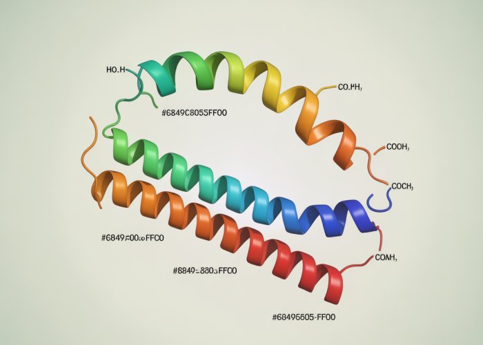
Primary Structure’s Orchestration of Protein Folding
The primary structure of a protein, the linear sequence of amino acids, serves as the blueprint that dictates all subsequent levels of structural organization: secondary, tertiary, and quaternary. This sequence isn’t just a random assortment; it’s a meticulously encoded message that guides the protein towards its functional conformation.
From Sequence to Structure: A Hierarchical Process
The journey from a linear chain of amino acids to a functional three-dimensional protein is a complex and fascinating process, governed by the inherent properties of the amino acids themselves.
The primary structure lays the foundation for the formation of secondary structures, such as alpha-helices and beta-sheets. These local, repeating structures arise from hydrogen bonds between the amino and carboxyl groups of amino acids in the polypeptide backbone.
The specific sequence of amino acids determines where these secondary structural elements form and how they interact with each other.
The Amino Acid Sequence as a Folding Code
The amino acid sequence profoundly influences protein folding. Hydrophobic amino acids tend to cluster in the protein’s interior, away from the aqueous environment, while hydrophilic amino acids are more likely to be found on the surface, interacting with water molecules.
Charged amino acids can form salt bridges, contributing to the protein’s stability, while cysteine residues can form disulfide bonds, cross-linking different parts of the polypeptide chain.
These interactions, dictated by the primary structure, drive the protein to fold into its unique three-dimensional tertiary structure, which is essential for its biological activity.
Incorrect folding, often caused by mutations in the amino acid sequence, can lead to non-functional proteins or even the formation of toxic aggregates, as seen in diseases like Alzheimer’s and Parkinson’s.
The Genetic Code: Translating DNA into Protein Sequence
The link between the genetic code and the primary structure is fundamental to understanding protein synthesis. DNA contains the instructions for building proteins, with each three-nucleotide codon specifying a particular amino acid.
This genetic information is transcribed into messenger RNA (mRNA), which then serves as a template for protein synthesis on ribosomes.
During translation, transfer RNA (tRNA) molecules, each carrying a specific amino acid, recognize the codons on the mRNA and deliver the corresponding amino acids to the growing polypeptide chain.
The sequence of codons in the mRNA directly determines the sequence of amino acids in the protein, highlighting the central role of the genetic code in dictating protein primary structure and, consequently, its function.
Charged amino acids can form salt bridges, further stabilizing the folded structure. Disulfide bonds between cysteine residues can also contribute to the protein’s stability. Understanding these interactions, guided by the primary sequence, is crucial for predicting a protein’s final conformation.
Implications and Applications: Why Primary Structure Matters
The determination of a protein’s primary structure transcends mere academic curiosity; it serves as a cornerstone for a myriad of applications with profound implications for medicine, biotechnology, and beyond. From predicting protein function to designing novel therapeutics, the information encoded within a protein’s amino acid sequence unlocks a wealth of possibilities.
Predicting Protein Function from Sequence
The primary sequence of a protein provides the most immediate clues regarding its function. By comparing a newly determined sequence to those of well-characterized proteins in databases, researchers can infer potential functions based on sequence homology. Conserved sequence motifs, often associated with specific enzymatic activities or binding domains, offer valuable insights into a protein’s role within a cell or organism.
For example, the presence of an ATP-binding motif strongly suggests that the protein is an enzyme that utilizes ATP as an energy source. Similarly, sequences resembling known DNA-binding domains implicate the protein in gene regulation. These predictions, while not definitive, provide a crucial starting point for experimental investigations into protein function.
Designing Novel Proteins and Peptides
The ability to manipulate protein primary structure has opened up new avenues for designing proteins with tailored properties and functions. Rational protein design leverages our understanding of the relationship between sequence and structure to create proteins with enhanced stability, altered substrate specificity, or novel binding capabilities.
This approach involves carefully selecting and arranging amino acids to achieve a desired three-dimensional conformation and, consequently, a specific function. Such de novo protein design holds immense promise for creating novel enzymes, biosensors, and therapeutic agents.
Understanding Disease Mechanisms Through Sequence Analysis
Mutations in the DNA sequence can lead to alterations in the amino acid sequence of a protein, potentially disrupting its structure and function. Understanding how these mutations affect protein behavior is crucial for elucidating the molecular basis of many diseases.
For example, in sickle cell anemia, a single amino acid substitution in the beta-globin chain of hemoglobin leads to the aggregation of hemoglobin molecules, resulting in the characteristic sickle shape of red blood cells. By identifying and characterizing such mutations, researchers can gain insights into the pathogenesis of diseases and develop targeted therapies.
Developing Targeted Therapies Based on Primary Structure
The primary structure of a protein can serve as a target for therapeutic intervention. Antibodies, for instance, can be designed to specifically recognize and bind to a particular region of a protein, inhibiting its function or marking it for destruction.
Moreover, small molecule drugs can be developed to interact with specific amino acid residues within a protein’s active site, thereby blocking its enzymatic activity. The design of such targeted therapies requires a detailed understanding of the protein’s primary structure and its three-dimensional conformation.
In cancer therapy, for example, many drugs target specific protein kinases, enzymes that play a crucial role in cell growth and proliferation. By inhibiting the activity of these kinases, these drugs can selectively kill cancer cells while sparing normal cells. The primary sequence of these kinases provides essential information for the design and optimization of these inhibitors.
Frequently Asked Questions About Protein Primary Structure
This section answers common questions about protein primary structure to help you understand this fundamental concept in biochemistry.
What exactly is a protein’s primary structure?
The primary structure of a protein refers to the linear sequence of amino acids that make up the polypeptide chain. This sequence is held together by peptide bonds, and it dictates the protein’s unique identity and ultimately, its function. Think of it as the specific order of letters that spell out a word.
How is the primary structure of a protein determined?
The protein’s primary structure is determined by the DNA sequence of the gene that encodes the protein. During protein synthesis, the ribosome reads the mRNA code (transcribed from the DNA) and assembles the amino acids in the precise order specified. This is the fundamental process that ensures the fidelity of proteins primary structure.
Why is knowing the proteins primary structure important?
Understanding the proteins primary structure is crucial because it dictates all higher levels of protein structure – secondary, tertiary, and quaternary. Changes in the amino acid sequence can drastically alter a protein’s shape and function, potentially leading to diseases or loss of function.
Can the primary structure of a protein be modified after synthesis?
Yes, the proteins primary structure can be modified after translation through post-translational modifications. These modifications, such as phosphorylation or glycosylation, can alter the protein’s activity, localization, or interactions with other molecules. However, they don’t change the underlying amino acid sequence itself.
So, now you’re a little more fluent in the language of proteins primary structure! Hopefully, you found this helpful and can impress your friends at the next biology hangout. Keep exploring, and who knows what protein secrets you’ll uncover next!