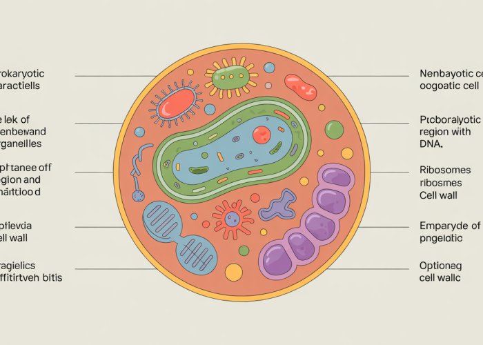
Prokaryotic cell characteristics define fundamental aspects of cellular biology, influencing diverse fields from microbial ecology to biotechnology. Ribosomes, essential cellular structures, exhibit notable differences in prokaryotes compared to eukaryotes, impacting protein synthesis efficiency. Investigating prokaryotic cell characteristics provides insights into the mechanisms used by entities like the Pasteur Institute, renowned for its contributions to understanding infectious diseases. The composition of the cell wall significantly differentiates prokaryotic cell characteristics from eukaryotic cells, contributing to their survival in various environments.
Prokaryotic cells represent a foundational element of life, a testament to simplicity and resilience. These microscopic entities, though often overlooked, are the architects of our planet’s ecosystems and play pivotal roles in processes ranging from nutrient cycling to human health.
In essence, they laid the groundwork for the evolution of all other life forms. To understand the intricacies of biology, medicine, and biotechnology, one must first grasp the fundamental principles governing the prokaryotic world.
Defining the Prokaryotic Cell
The term "prokaryote" itself holds a clue to its nature. Derived from the Greek words "pro" (before) and "karyon" (nucleus), it signifies a cell that existed before the evolution of the nucleus.
This lack of a membrane-bound nucleus is the defining characteristic that distinguishes prokaryotes from their more complex eukaryotic counterparts.
Prokaryotic cells are generally smaller and simpler in structure than eukaryotic cells. Their genetic material, typically a single circular chromosome, resides in a region called the nucleoid, rather than being enclosed within a nuclear membrane.
The Domains of Prokaryotes: Bacteria and Archaea
The prokaryotic world is divided into two distinct domains: Bacteria and Archaea. While both share the fundamental characteristics of prokaryotic cells, they differ significantly in their evolutionary history, genetic makeup, and biochemical pathways.
Bacteria are the more familiar of the two, encompassing a vast array of species that inhabit diverse environments, from soil and water to the human gut. They play crucial roles in nutrient cycling, decomposition, and various industrial processes.
Archaea, initially considered a subgroup of bacteria, are now recognized as a separate domain with unique characteristics. They are often found in extreme environments, such as hot springs, salt lakes, and anaerobic sediments.
Archaea’s unique adaptations to extreme conditions provide insights into the limits of life and offer potential applications in biotechnology.
Evolutionary Significance: The First Life Forms
Prokaryotes hold a special place in the history of life. They are believed to be the first organisms to inhabit Earth, emerging billions of years ago. Their simple structure and metabolic versatility allowed them to thrive in the harsh conditions of the early Earth.
Through photosynthesis, cyanobacteria, a type of bacteria, oxygenated the atmosphere, paving the way for the evolution of more complex, oxygen-dependent life forms.
The evolutionary journey of prokaryotes has shaped the trajectory of life on Earth.
The Importance of Studying Prokaryotes
Understanding prokaryotes is paramount in numerous scientific disciplines.
In microbiology, they are essential model organisms for studying fundamental biological processes, such as DNA replication, protein synthesis, and metabolic pathways.
In medicine, prokaryotes are both allies and adversaries. Many bacteria are beneficial, contributing to digestion and immunity, while others are pathogens that cause infectious diseases. The study of prokaryotes is crucial for developing new antibiotics and vaccines.
In biotechnology, prokaryotes are harnessed for a wide range of applications, including the production of pharmaceuticals, biofuels, and enzymes. Genetic engineering of prokaryotes has revolutionized the production of insulin and other life-saving drugs.
The continued exploration of the prokaryotic world promises to unlock new insights into the nature of life and provide innovative solutions to global challenges.
Prokaryotic cells represent a foundational element of life, a testament to simplicity and resilience. These microscopic entities, though often overlooked, are the architects of our planet’s ecosystems and play pivotal roles in processes ranging from nutrient cycling to human health.
In essence, they laid the groundwork for the evolution of all other life forms. To understand the intricacies of biology, medicine, and biotechnology, one must first grasp the fundamental principles governing the prokaryotic world. This brings us to one of the most defining features of these cells: the absence of a true nucleus. How do prokaryotes manage their genetic material without this dedicated compartment? Let’s explore the fascinating world of the nucleoid.
No Nucleus, No Problem: The Simplicity of the Nucleoid
The hallmark of a prokaryotic cell is, undoubtedly, the absence of a membrane-bound nucleus. Unlike eukaryotic cells, which carefully sequester their DNA within a nucleus, prokaryotes take a different approach.
Instead of a nucleus, they possess a region called the nucleoid. This is where the cell’s genetic material resides.
Absence of a True Nucleus
It’s crucial to emphasize that the nucleoid isn’t simply a less organized nucleus. It lacks the nuclear envelope, the double membrane that defines the eukaryotic nucleus.
Moreover, prokaryotic cells lack other membrane-bound organelles like mitochondria, the endoplasmic reticulum, and the Golgi apparatus.
This absence of internal compartmentalization reflects the simplicity of prokaryotic cell structure.
The Nucleoid Region
Within the nucleoid, you’ll find the bacterial chromosome, which is typically a single, circular DNA molecule.
This chromosome contains the vast majority of the genes necessary for the cell’s survival and function. It’s important to note, however, that the nucleoid is not just a bag of DNA.
Proteins and RNA molecules are actively involved in organizing and regulating the DNA.
DNA Organization and Compaction
Given the relatively small size of a prokaryotic cell, a significant challenge is how to fit a long DNA molecule within the confines of the nucleoid.
The solution lies in a process called supercoiling. The DNA is twisted and folded upon itself, creating a compact structure.
Think of it like tightly winding a rubber band to make it smaller. This supercoiling is facilitated by enzymes called topoisomerases, which can introduce or remove twists in the DNA.
This allows for efficient packaging of the genetic material.
The Role of Plasmids
In addition to the main chromosome, many prokaryotes also contain plasmids. These are small, circular DNA molecules that are separate from the chromosome and capable of independent replication.
Plasmids often carry genes that provide additional benefits to the cell, such as antibiotic resistance or the ability to metabolize specific compounds.
They can be transferred between bacteria through horizontal gene transfer. This contributes to genetic diversity and adaptation.
Nucleoid vs. Eukaryotic Nucleus
The contrast between the nucleoid and the eukaryotic nucleus is stark. The eukaryotic nucleus is a highly organized structure with a defined boundary, the nuclear envelope.
Within the nucleus, the DNA is linear and organized into chromosomes, which are further packaged with histone proteins into chromatin.
The nucleus also contains a nucleolus, a region where ribosomes are assembled. The nucleoid, on the other hand, is a more dynamic and less structurally defined region.
Its organization relies on supercoiling and associated proteins, but lacks the complex architecture of the eukaryotic nucleus. This structural difference is fundamental to understanding the differences in gene regulation and cell division between prokaryotes and eukaryotes.
No Nucleus, No Problem: The Simplicity of the Nucleoid, while a defining characteristic, isn’t the only structural feature that sets prokaryotes apart. While the nucleoid houses the genetic material, prokaryotic cells also require a robust outer layer to maintain their shape, withstand internal pressure, and interact with their environment. This brings us to the cell wall, a structure as crucial as it is varied, especially when differentiating between bacteria and archaea.
The Protective Armor: Cell Wall Structure and Function
The cell wall is an indispensable structure found in nearly all prokaryotic cells, serving as a rigid, protective barrier that lies outside the cell membrane. Its primary function is to provide structural support and protect the cell from osmotic pressure, which can be significant due to the high concentration of solutes inside the cell. Without a cell wall, many prokaryotes would simply burst in a hypotonic environment.
Bacterial Cell Walls: The Peptidoglycan Fortress
The bacterial cell wall is primarily composed of peptidoglycan, a unique polymer consisting of sugar and amino acid chains. These chains are cross-linked, forming a mesh-like structure that encases the entire cell.
Think of it as a molecular chain-mail, providing strength and rigidity. The specific composition and thickness of the peptidoglycan layer can vary between different bacterial species, contributing to their distinct characteristics.
Gram Staining: A Key to Bacterial Identification
One of the most important techniques in microbiology is the Gram stain, which differentiates bacteria based on their cell wall structure. This technique, developed by Hans Christian Gram, categorizes bacteria as either Gram-positive or Gram-negative.
Gram-positive bacteria have a thick peptidoglycan layer that retains the crystal violet stain, resulting in a purple color under the microscope. Gram-negative bacteria, on the other hand, have a thinner peptidoglycan layer and an outer membrane containing lipopolysaccharide (LPS).
During the Gram staining procedure, the crystal violet is easily washed away from Gram-negative cells, which are then counterstained with safranin, resulting in a pink or red color.
Structural Differences and Staining Variations
The key to the differential staining lies in the structure of the cell wall. The thick peptidoglycan layer of Gram-positive bacteria acts like a sponge, trapping the crystal violet-iodine complex and preventing it from being washed away during the decolorization step.
In contrast, the thin peptidoglycan layer of Gram-negative bacteria is unable to retain the crystal violet complex. The outer membrane, unique to Gram-negative bacteria, also plays a role by hindering the penetration of the crystal violet.
Lipopolysaccharide (LPS) and Endotoxin Properties
The outer membrane of Gram-negative bacteria contains lipopolysaccharide (LPS), also known as endotoxin. LPS is a potent immunostimulant that can trigger a strong inflammatory response in animals.
Infections with Gram-negative bacteria can lead to the release of LPS, causing fever, shock, and even death. The endotoxic properties of LPS make Gram-negative bacteria a significant concern in healthcare settings.
Archaeal Cell Walls: Beyond Peptidoglycan
While bacteria rely on peptidoglycan, archaea exhibit a greater diversity in cell wall composition. Many archaea possess a cell wall made of pseudopeptidoglycan, which is similar to peptidoglycan but differs in its chemical structure.
Other archaea may have cell walls composed of various polysaccharides, proteins, or glycoproteins. The diversity in archaeal cell wall composition reflects their adaptation to a wide range of extreme environments. For example, some archaea thrive in hot springs, while others inhabit highly saline or acidic environments. Their unique cell walls provide the necessary protection to survive in these harsh conditions.
Movement and Attachment: Flagella, Pili, and Capsules
Beyond the rigid protection offered by the cell wall, prokaryotic cells possess other specialized structures that enable them to navigate their environment, adhere to surfaces, and evade host defenses. These structures—flagella, pili (fimbriae), and capsules—are crucial for survival and, in many cases, contribute significantly to the pathogenicity of disease-causing bacteria. Understanding their structure and function is paramount to comprehending prokaryotic behavior and developing strategies to combat bacterial infections.
Flagella: Propellers of the Prokaryotic World
Flagella are whip-like appendages that provide motility for many prokaryotic cells. These intricate structures extend from the cell surface and rotate to propel the cell through liquid environments. Unlike eukaryotic flagella, which move in a wave-like motion, prokaryotic flagella rotate like a propeller.
This rotation is powered by a molecular motor located at the base of the flagellum, driven by the flow of ions (either protons or sodium ions) across the cell membrane.
Flagellar Arrangements
The arrangement of flagella can vary significantly among different bacterial species, leading to distinct patterns of motility. These arrangements are often used as a distinguishing characteristic in bacterial identification.
-
Monotrichous: A single flagellum extends from one pole of the cell.
-
Amphitrichous: A single flagellum extends from both poles of the cell.
-
Lophotrichous: A tuft of flagella extends from one or both poles of the cell.
-
Peritrichous: Flagella are distributed over the entire surface of the cell.
The Mechanism of Flagellar Rotation
The bacterial flagellum is a remarkable feat of biological engineering. Its rotation is driven by a complex motor embedded in the cell membrane and cell wall. This motor consists of several proteins that interact to generate torque. The flow of protons (H+) or sodium ions (Na+) across the membrane provides the energy for this rotation.
The flagellar motor can rotate at incredibly high speeds, allowing bacteria to move rapidly through their environment. Bacteria can also control the direction of flagellar rotation, allowing them to swim towards attractants (chemotaxis) or away from repellents.
Pili (Fimbriae): Anchors to the Environment
Pili, also known as fimbriae, are short, hair-like appendages that extend from the surface of many prokaryotic cells. Unlike flagella, pili are not involved in motility. Instead, they primarily function in attachment to surfaces, including host cells, inert surfaces, and other bacteria.
Pili are composed of protein subunits called pilins, which assemble to form a thin, flexible rod. The tip of the pilus often contains specific adhesins that bind to complementary receptors on the target surface.
This specific binding is crucial for bacterial colonization and the formation of biofilms.
Capsules: Slippery Shields
Capsules are a layer of polysaccharide or protein that surrounds the cell wall of some prokaryotic cells. This layer is typically sticky and can provide several advantages to the bacterium.
One of the most important functions of the capsule is to protect the cell from phagocytosis by immune cells. The capsule’s slippery surface makes it difficult for phagocytes to engulf and destroy the bacterium.
In addition to protection, capsules can also facilitate adherence to surfaces, contributing to biofilm formation and colonization. The sticky nature of the capsule allows bacteria to adhere more effectively to host tissues or other surfaces in their environment.
Ribosomes and Protein Synthesis: The Workhorses of the Cell
Beyond movement and protection, prokaryotic cells must also synthesize the proteins necessary for their survival and function. This critical task is carried out by ribosomes, intricate molecular machines that translate genetic information into functional proteins.
The Structure of Prokaryotic Ribosomes
Prokaryotic ribosomes, while sharing the same fundamental function as their eukaryotic counterparts, possess a distinct structure. These ribosomes are classified as 70S ribosomes, where "S" stands for Svedberg units, a measure of sedimentation rate during centrifugation, reflecting size and shape.
The 70S ribosome is composed of two subunits: a large 50S subunit and a small 30S subunit. Each subunit consists of ribosomal RNA (rRNA) molecules and ribosomal proteins.
The 30S subunit is responsible for binding to messenger RNA (mRNA), the template carrying the genetic code.
The 50S subunit catalyzes the formation of peptide bonds, linking amino acids together to form the growing polypeptide chain.
Ribosomes: Translating the Genetic Code
The primary role of ribosomes is to translate the information encoded in mRNA into a specific sequence of amino acids, thereby creating a protein.
This process, known as translation, occurs in a series of precisely coordinated steps. The ribosome binds to the mRNA and moves along it, reading the genetic code in triplets called codons.
Each codon specifies a particular amino acid, which is brought to the ribosome by a transfer RNA (tRNA) molecule. The ribosome then links the amino acids together, forming a polypeptide chain that will eventually fold into a functional protein.
Prokaryotic vs. Eukaryotic Ribosomes: A Key Difference
A crucial difference exists between prokaryotic and eukaryotic ribosomes. Eukaryotic cells possess 80S ribosomes, which are larger and more complex than the 70S ribosomes found in prokaryotes.
This structural difference is not merely academic; it has significant implications for medicine.
Many antibiotics specifically target the 70S ribosomes of bacteria, inhibiting protein synthesis without affecting the host’s eukaryotic ribosomes. This selective toxicity is what makes these antibiotics effective in treating bacterial infections.
Drugs like tetracycline and streptomycin, for instance, bind to the 30S subunit, while others like erythromycin bind to the 50S subunit, disrupting the translation process.
A Glimpse into Protein Synthesis
Protein synthesis is a complex and highly regulated process. It involves a series of steps, from the initiation of translation to the termination and release of the newly synthesized protein.
Briefly, the process involves:
- Initiation: The ribosome binds to the mRNA and begins scanning for the start codon.
- Elongation: tRNA molecules bring amino acids to the ribosome, which adds them to the growing polypeptide chain.
- Termination: The ribosome encounters a stop codon, signaling the end of translation. The polypeptide chain is released and folds into its functional shape.
Understanding the intricacies of protein synthesis is crucial for comprehending how prokaryotic cells function and how we can develop effective strategies to combat bacterial infections.
Small but Mighty: Size, Metabolism, and Reproduction
Having explored the inner workings of protein synthesis, it’s time to appreciate the overall scale and life cycle of these microscopic powerhouses. Despite their diminutive size, prokaryotic cells exhibit an astonishing range of metabolic strategies and reproductive capabilities, enabling them to thrive in virtually every environment on Earth.
Size Matters: The World of Micrometers
Prokaryotic cells are generally much smaller than their eukaryotic counterparts.
Their size typically ranges from 0.5 to 5 micrometers (μm) in diameter.
This small size has profound implications for their surface area-to-volume ratio, facilitating rapid nutrient uptake and waste removal.
This efficiency contributes to their rapid growth rates and ability to quickly adapt to changing environmental conditions.
Metabolic Versatility: Masters of Energy Acquisition
One of the most remarkable features of prokaryotes is their diverse metabolic capabilities. Unlike eukaryotes, which are largely limited to aerobic respiration and fermentation, prokaryotes have evolved a vast array of strategies for obtaining energy and nutrients.
Autotrophs are able to synthesize their own organic compounds from inorganic sources, such as carbon dioxide.
Photoautotrophs, like cyanobacteria, use sunlight as their energy source, while chemoautotrophs obtain energy from chemical reactions involving inorganic compounds like sulfur or iron.
Heterotrophs, on the other hand, obtain their energy and carbon from organic compounds produced by other organisms.
This group includes a wide range of bacteria and archaea that can utilize diverse carbon sources, from simple sugars to complex polymers.
Some prokaryotes are even capable of chemoheterotrophy, obtaining both energy and carbon from chemical compounds.
This metabolic versatility allows prokaryotes to occupy a wide range of ecological niches and play crucial roles in nutrient cycling and biogeochemical processes.
Reproduction: The Speed of Binary Fission
Prokaryotes primarily reproduce asexually through a process called binary fission.
This relatively simple process involves the replication of the cell’s DNA, followed by cell division, resulting in two identical daughter cells.
Binary fission is a rapid and efficient mode of reproduction, allowing prokaryotic populations to double in as little as 20 minutes under optimal conditions.
This rapid growth rate enables them to quickly colonize new environments and outcompete other organisms.
Horizontal Gene Transfer: A Shortcut to Evolution
While binary fission produces genetically identical offspring, prokaryotes also have mechanisms for exchanging genetic material horizontally, bypassing traditional inheritance.
This horizontal gene transfer (HGT) allows them to acquire new genes from unrelated organisms, leading to rapid adaptation and evolution.
There are three main mechanisms of HGT:
- Conjugation: Direct transfer of DNA between two cells through a physical connection (pilus).
- Transduction: Transfer of DNA via a bacteriophage (virus that infects bacteria).
- Transformation: Uptake of free DNA from the environment.
HGT plays a crucial role in the spread of antibiotic resistance genes among bacteria, posing a significant challenge to public health.
It also contributes to the evolution of new metabolic capabilities and the adaptation of prokaryotes to diverse environments.
FAQs About Prokaryotic Cell Characteristics
This FAQ section addresses common questions about prokaryotic cell characteristics to help you better understand these simple yet vital cells.
What are the 5 key things to know about prokaryotic cells?
The five key things to know are that prokaryotic cells lack a nucleus, have simple internal structures, reproduce asexually, are small, and have diverse metabolic capabilities. These prokaryotic cell characteristics define their fundamental nature.
How does not having a nucleus impact prokaryotic cells?
The absence of a nucleus means that the genetic material (DNA) in prokaryotic cells isn’t enclosed within a membrane. It resides in a region called the nucleoid. This affects how prokaryotic cell characteristics like DNA replication and gene expression are regulated.
What are some examples of prokaryotic organisms?
Bacteria and Archaea are the two main domains of life that are composed of prokaryotic cells. These organisms exhibit all the typical prokaryotic cell characteristics.
How do prokaryotic cell characteristics affect their ability to adapt?
The small size, simple structure, and diverse metabolic capabilities of prokaryotic cells enable them to adapt rapidly to changing environments. Their asexual reproduction also allows for quick propagation of beneficial mutations, making prokaryotic cell characteristics crucial for their survival and evolution.
So, there you have it – a quick look at prokaryotic cell characteristics! Hopefully, this gave you a clearer picture. Now go forth and explore the amazing world of these tiny but mighty cells!