Cellular biology employs centrifugation, a powerful separation technique. This technique, integral to understanding cellular components, produces a pellet. Pellet definition biology hinges on understanding its relationship to supernatant. Specifically, a cell fraction precipitates forming the pellet, while the liquid supernatant contains other cellular constituents. Understanding pellet definition biology requires a clear understanding of both the cellular components and the process of cell fractionation. In the context of cell fractionation, where the separation of organelles like mitochondria happens, pellet definition biology means the isolated organelles within the solid precipitate at the bottom of the centrifuge tube are defined and characterized. This understanding is fundamental in various research applications.
In the realm of biological research, seemingly simple components can hold profound significance. Among these is the pellet, a concentrated mass of cellular material obtained through centrifugation. Understanding what pellets are, how they are formed, and what they contain is paramount for a diverse range of scientific investigations.
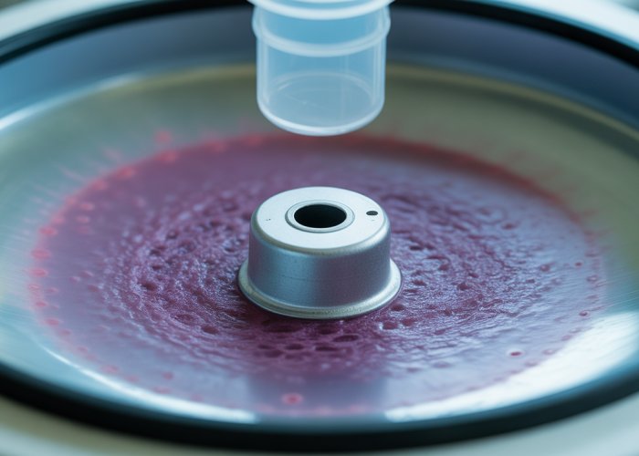
Defining the Biological Pellet
In biological terms, a pellet refers to the solid fraction that sediments to the bottom of a tube after centrifugation. This process involves spinning a liquid sample at high speeds, causing denser components to separate from the solution and form a compacted mass, the pellet, while less dense components remain in the supernatant (the liquid above the pellet).
The composition of a pellet can vary widely depending on the initial sample and the centrifugation parameters employed, but it commonly includes cells, cell fragments, organelles, proteins, and nucleic acids.
Why Pellets Matter: Their Importance in Research
Pellets are not merely waste products or byproducts of experimental procedures; they are valuable sources of biological information. The ability to isolate and analyze specific cellular components within a pellet allows researchers to:
-
Study cellular processes: By examining the proteins, organelles, or other molecules concentrated in a pellet, scientists can gain insights into the biochemical pathways and molecular mechanisms that govern cellular function.
-
Purify specific components: Pellets can be used to isolate and purify specific organelles, proteins, or other biomolecules of interest for further study.
-
Diagnose diseases: Analyzing the composition of pellets obtained from patient samples can aid in the diagnosis and monitoring of various diseases.
-
Develop new therapies: Pellets are crucial in the development of new therapies, offering insights into drug targets and mechanisms of action.
Scope of This Guide: A Comprehensive Overview
This guide aims to provide a comprehensive understanding of pellets in biological research. We will explore the fundamental principles behind pellet formation, including cell fractionation and centrifugation techniques. We will delve into advanced methods like differential and ultracentrifugation, which allow for the isolation of pellets with specific compositions.
Furthermore, we will discuss the typical components found in pellets, such as organelles and proteins, and explore methods for analyzing their presence and abundance. Finally, we will examine the diverse applications of pellets in various biological research areas, including the study of cellular proteins and the isolation of organelles. By the end of this guide, readers will have a solid foundation for understanding the significance of pellets and their applications in advancing biological knowledge.
In essence, pellets become reservoirs of concentrated biological information, enabling researchers to dissect complex cellular processes, purify specific components, and even diagnose diseases. But how exactly are these valuable pellets formed? The answer lies in mastering the foundational techniques of cell fractionation and centrifugation.
Cell Fractionation and Centrifugation: The Foundational Techniques
Cell fractionation and centrifugation are indispensable techniques in biological research, serving as the bedrock for isolating and studying cellular components. These processes, working in tandem, allow scientists to separate complex mixtures of cellular material into distinct fractions, ultimately leading to the formation of pellets containing specific components of interest.
Cell Fractionation: Dissecting the Cellular Landscape
Cell fractionation is the process of separating cellular components while preserving the individual functions of each component.
It is a crucial initial step in many biochemical and cell biology experiments.
The primary objective of cell fractionation is to isolate organelles and other subcellular structures from a complex mixture, allowing for their individual study.
This process typically involves a series of steps, starting with cell lysis to break open the cells and release their contents.
The resulting homogenate, a mixture of cellular components, is then subjected to differential centrifugation, as we’ll explore later.
The significance of cell fractionation in pellet isolation cannot be overstated. It sets the stage for subsequent centrifugation steps by creating a heterogeneous mixture ready for separation based on size, shape, and density.
Without effective cell fractionation, the resulting pellets would be a jumbled mess of everything, hindering meaningful analysis.
Centrifugation: The Core Process of Pellet Formation
Centrifugation is the workhorse technique driving pellet formation.
It leverages centrifugal force to separate components based on their mass, size, and density.
The process involves spinning a sample at high speeds within a centrifuge, generating a centrifugal force that causes denser components to sediment towards the bottom of the tube, forming the pellet.
Lighter components remain suspended in the liquid above the pellet, known as the supernatant.
The speed and duration of centrifugation are critical parameters that determine which components will be pelleted.
Higher speeds and longer durations result in the sedimentation of smaller and less dense components.
Centrifugation is directly responsible for the creation of pellets.
By carefully controlling the centrifugation parameters, researchers can selectively pellet specific cellular components, such as nuclei, mitochondria, or ribosomes.
This level of control is essential for purifying and studying individual organelles or macromolecules.
Key Terms in Pellet Formation: Sedimentation and Supernatant
Understanding the terminology associated with pellet formation is crucial for accurate interpretation of experimental results.
Sedimentation: The Driving Force
Sedimentation refers to the process by which particles settle out of a suspension under the influence of gravity or, more commonly in the context of cell biology, centrifugal force.
During centrifugation, the centrifugal force causes denser particles to move through the liquid medium and accumulate at the bottom of the tube.
The rate of sedimentation depends on several factors, including the size, shape, and density of the particles, as well as the viscosity of the medium and the centrifugal force applied.
Sedimentation is the fundamental mechanism underlying pellet formation.
Supernatant: The Liquid Above
The supernatant is the liquid fraction that remains above the pellet after centrifugation.
It contains the lighter, less dense components that did not sediment under the applied centrifugal force.
The composition of the supernatant is inversely related to the composition of the pellet.
If the pellet contains primarily nuclei, the supernatant will contain primarily cytosolic proteins and smaller organelles.
The supernatant is not merely a waste product; it can be a valuable source of information.
It often contains soluble proteins, enzymes, and other molecules of interest that can be further analyzed or purified.
Cell fractionation lays the groundwork, separating cellular components based on gross properties. But to achieve finer, more specific separations and ultimately isolate pellets of distinct compositions, more sophisticated centrifugation techniques are required. This is where differential and ultracentrifugation come into play.
Differential and Ultracentrifugation: Advanced Pellet Isolation
Differential and ultracentrifugation represent the next tier in pellet isolation techniques, offering enhanced precision in separating cellular components. These methods allow researchers to isolate specific organelles and macromolecules with greater accuracy than basic centrifugation alone.
Differential Centrifugation: Separating by Size and Density
Differential centrifugation is a sequential centrifugation process that leverages varying speeds and durations to separate cellular components based on their size and density. It’s a workhorse technique in cell biology, providing a relatively simple and scalable method for initial fractionation.
The Process of Differential Centrifugation
The process begins with a cell homogenate, obtained after cell lysis and initial fractionation. This homogenate is then subjected to a series of centrifugation steps, each with increasing speeds. At each speed, components of a certain size and density will pellet, while smaller, less dense components remain in the supernatant.
The supernatant is then carefully removed and subjected to the next centrifugation step at a higher speed. This process is repeated several times, resulting in a series of pellets, each enriched in different cellular components.
For example, a low-speed spin might pellet nuclei and whole cells, while subsequent higher-speed spins might pellet mitochondria, lysosomes, and finally, ribosomes and endoplasmic reticulum fragments.
Impact on Pellet Composition
Differential centrifugation significantly impacts pellet composition. By carefully controlling the centrifugation parameters (speed and time), researchers can enrich for specific organelles or cellular fractions in each pellet.
The initial low-speed spins yield pellets containing larger, denser structures. Progressively higher speeds yield pellets containing smaller and less dense components. This allows for a relatively targeted separation of cellular constituents.
However, it’s important to note that differential centrifugation rarely achieves perfect separation. Pellets often contain a mixture of components due to overlapping sedimentation rates. Therefore, further purification steps may be required depending on the desired level of purity.
Ultracentrifugation: When and How to Use
Ultracentrifugation takes centrifugation to the extreme, employing extremely high speeds to separate even the smallest cellular components, such as viruses, macromolecules, and even individual proteins.
This technique requires specialized equipment capable of generating immense centrifugal forces.
Understanding the Application of Ultracentrifugation
Ultracentrifugation is indispensable when dealing with very small particles or when high resolution separation is required. Its applications range from isolating specific proteins or nucleic acids to purifying viral particles for vaccine development or structural studies.
It is also used to separate different types of membrane vesicles, liposomes, and other nanoscale structures. Due to its high resolving power, ultracentrifugation is also useful for separating macromolecules based on subtle differences in density through density gradient centrifugation.
Advanced Isolation Techniques using Ultracentrifugation
Ultracentrifugation enables several advanced isolation techniques, including rate-zonal centrifugation and isopycnic centrifugation (also known as equilibrium density gradient centrifugation).
Rate-zonal centrifugation separates particles based on size and shape. The sample is layered on top of a density gradient, and during centrifugation, particles migrate through the gradient at different rates depending on their size and shape.
Isopycnic centrifugation, on the other hand, separates particles based on their buoyant density. The sample is mixed with a density gradient, and during centrifugation, particles migrate to the point in the gradient where their density matches the density of the surrounding medium.
These advanced techniques, though more complex, provide unparalleled resolution in separating cellular components, leading to highly purified pellets suitable for in-depth analysis.
Analyzing Pellet Composition: Organelles and Proteins
The isolation of a pellet is only the first step. To truly unlock its potential, researchers must delve into its composition, identifying the specific organelles and proteins it contains. This analysis is crucial for verifying the success of the fractionation process and for interpreting experimental results in a meaningful way.
Organelles in Pellets: Identifying Common Components
Pellets are rarely composed of a single, homogenous substance. Instead, they typically contain a mixture of cellular components, with the specific composition depending on the cell type, the fractionation technique used, and the experimental design. Identifying the organelles present within a pellet is therefore essential.
Which Organelles Are Typically Found in Pellets?
The organelles most commonly found in pellets include nuclei, mitochondria, lysosomes, peroxisomes, and fragments of the endoplasmic reticulum (ER) and Golgi apparatus.
-
Nuclei, being relatively large and dense, tend to pellet at lower centrifugation speeds.
-
Mitochondria and lysosomes usually pellet at intermediate speeds.
-
Ribosomes and fragments of the ER and Golgi often require higher speeds for sedimentation.
The presence of specific organelles can also be influenced by the disruption method used during cell lysis. Harsh methods may fragment organelles, leading to their presence in pellets obtained at higher centrifugation speeds than expected.
Correlating the Presence of Specific Organelles to the Experimental Goal
The identification of organelles within a pellet should always be considered in the context of the experimental goal. For example, if the goal is to isolate pure mitochondria for the study of oxidative phosphorylation, the pellet obtained at the mitochondrial fractionation step should be carefully analyzed to confirm the enrichment of mitochondria and the absence of other contaminating organelles.
Techniques like electron microscopy can provide a visual confirmation of organelle identity and purity. Similarly, Western blotting using antibodies specific to marker proteins for different organelles can be used to quantify the relative abundance of each organelle within the pellet.
Proteins in Pellets: A Major Component
In addition to organelles, pellets are also rich in proteins. These proteins can be integral membrane proteins of organelles, soluble proteins trapped within organelles, or proteins that have aggregated during the fractionation process. Analyzing the protein content of a pellet is crucial for understanding its function and for identifying potential contaminants.
Discussing Common Proteins Found in Pellets
The specific proteins found in a pellet will depend on the organelles and cellular components present. For example, a mitochondrial pellet will be enriched in proteins involved in oxidative phosphorylation, while a nuclear pellet will be enriched in histones and transcription factors. Pellets may also contain cytoskeletal proteins, such as actin and tubulin, especially if the cell lysis procedure was not optimized.
Analyzing the protein composition can also reveal valuable information about cellular processes.
For example, the presence of specific signaling proteins in a pellet may indicate the activation of a particular signaling pathway.
Methods for Analyzing Protein Content in Pellets
Several methods can be used to analyze the protein content of pellets.
-
SDS-PAGE (sodium dodecyl sulfate-polyacrylamide gel electrophoresis), followed by staining with Coomassie blue or silver stain, allows for the visualization of the protein profile of the pellet.
-
Western blotting, using antibodies specific to target proteins, can be used to identify and quantify the abundance of specific proteins.
-
Mass spectrometry offers a powerful and comprehensive approach for identifying all the proteins present in a pellet. This technique can be used to generate a "proteomic fingerprint" of the pellet, providing a detailed snapshot of its protein composition.
Furthermore, enzyme activity assays can be performed on pellets to assess the activity of specific enzymes associated with particular organelles or cellular processes. This can be particularly useful for confirming the functional integrity of organelles isolated by cell fractionation.
Analyzing pellet composition allows researchers to identify the building blocks within: organelles and proteins. But this knowledge isn’t just for characterization; it unlocks a range of applications. Understanding the composition of pellets provides a foundation to explore the many ways they are used in biological research.
Applications of Pellets in Biological Research
Pellets, the concentrated products of cell fractionation and centrifugation, are far more than just byproducts. They represent enriched fractions of cellular components that enable focused investigation in a wide array of biological studies. Their applications span diverse areas, but are especially critical in the study of cellular proteins and organelle isolation.
Using Pellets in the Study of Cellular Proteins
Pellets provide a concentrated source of proteins, which is invaluable for a variety of proteomic analyses.
By isolating proteins within a pellet, researchers can reduce the complexity of their samples and focus on specific subsets of the proteome. This targeted approach is particularly useful when studying low-abundance proteins, which may be difficult to detect in whole-cell lysates.
Protein Purification and Characterization
One of the most common applications is protein purification. Pellets containing specific organelles or protein aggregates can be subjected to further purification steps, such as chromatography, to isolate individual proteins of interest.
The purified proteins can then be characterized using a variety of techniques, including:
-
Mass spectrometry to identify and quantify proteins.
-
Western blotting to confirm the presence and abundance of specific proteins.
-
Enzyme activity assays to measure the functional activity of enzymes.
Analyzing Protein Interactions
Pellets are also useful for studying protein-protein interactions. Co-immunoprecipitation (Co-IP) experiments, for example, can be performed using protein extracts from pellets to identify proteins that interact with a specific target protein.
The interacting proteins can then be identified by mass spectrometry, providing insights into the function and regulation of the target protein. Furthermore, the pelletized proteins can be examined by cross-linking strategies. Cross-linking stabilizes weak or transient interactions.
Studying Protein Aggregation
Protein aggregation is implicated in several neurodegenerative diseases, and pellets can be used to study the mechanisms underlying this process. By isolating protein aggregates from cell lysates or tissue samples, researchers can characterize their composition, structure, and toxicity.
These studies can provide insights into the pathogenesis of these diseases and identify potential therapeutic targets.
Applications in Isolating Organelles
Beyond protein studies, pellets serve as a critical starting point for isolating and studying specific organelles. The ability to enrich for a particular organelle within a pellet allows for detailed investigations into its structure, function, and role in cellular processes.
Mitochondria Isolation for Bioenergetics Studies
Mitochondria, the powerhouses of the cell, are frequently isolated from pellets. This allows researchers to investigate:
-
Oxidative phosphorylation: the process by which ATP is generated.
-
Mitochondrial dynamics: the fusion and fission of mitochondria.
-
Mitochondrial DNA (mtDNA): its structure and function.
By studying isolated mitochondria, researchers can gain a deeper understanding of mitochondrial function in both health and disease.
Lysosome Isolation for Degradation Studies
Lysosomes, the cell’s recycling centers, are also often isolated from pellets. This enables researchers to study:
-
Autophagy: the process by which cells degrade and recycle their own components.
-
Lysosomal storage disorders: genetic diseases caused by defects in lysosomal enzymes.
-
Drug delivery: using lysosomes as targets for drug delivery.
The use of pellets as a starting point ensures that the lysosomes are relatively free from other cellular components, which allows for more accurate and reliable results.
Studying Other Organelles
Pellets can also be used to isolate other organelles, such as:
-
Peroxisomes: involved in lipid metabolism and detoxification.
-
Endoplasmic reticulum (ER): involved in protein synthesis and folding.
-
Golgi apparatus: involved in protein processing and packaging.
The specific method used to isolate these organelles will depend on the experimental goal and the characteristics of the organelle of interest.
Frequently Asked Questions: Pellet Definition Biology
Here are some common questions regarding pellet definition biology and their role in various biological processes and experiments.
What exactly is a pellet in the context of biology?
In pellet definition biology, a pellet refers to the solid material that collects at the bottom of a tube after centrifugation. This sediment is typically composed of cells, cell debris, proteins, or other biological materials depending on the experiment.
How is a pellet formed during centrifugation?
During centrifugation, samples are spun at high speeds. The centrifugal force causes denser components in the sample to move outwards and settle at the bottom of the tube, forming a pellet. The less dense supernatant remains above the pellet.
What types of experiments use pellets?
Pellets are crucial in many experiments. For example, cell lysis protocols use centrifugation to separate cellular debris (pellet) from the desired protein or DNA in the supernatant. This is fundamental to pellet definition biology applications.
Why is it important to carefully handle a pellet after centrifugation?
The pellet often contains the key material of interest for downstream analysis. Disrupting or losing the pellet can compromise experimental results. Proper resuspension techniques are also important, ensuring the pellet is evenly distributed for further procedures.
So, now you’ve got a solid grasp on pellet definition biology! Go forth, explore the cellular world, and remember this guide whenever you’re dealing with cellular sediments. Happy centrifuging!