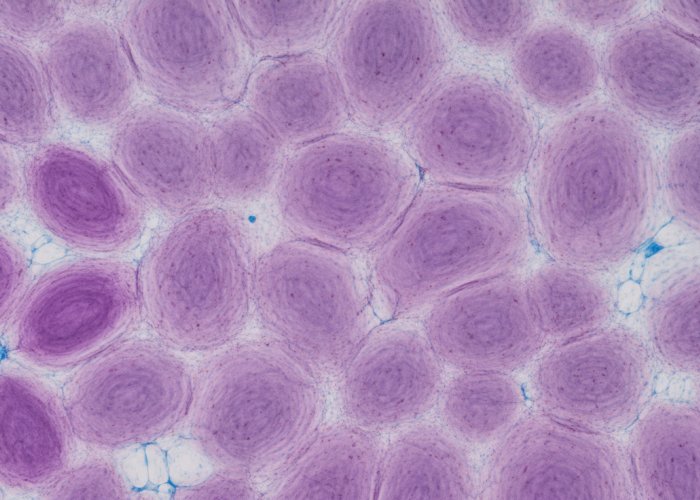
The fascinating world of onion epidermal cells, often explored using a light microscope, reveals a complex structure crucial for the plant’s protection. Plant biology students frequently study these cells to understand fundamental concepts of cellular organization. The Carolina Biological Supply Company is a common resource for obtaining prepared slides, facilitating this learning. Observing onion epidermal cells under magnification showcases the clear cell walls, a characteristic feature further explained by the work of Robert Hooke and other pioneering microscopists.

The world around us teems with hidden wonders, unseen universes contained within the smallest of spaces. To truly understand the intricacies of life, we must venture beyond what the naked eye can perceive and delve into the realm of the microscopic. Microscopy, the science of visualizing these minute structures, is therefore an indispensable tool in biological research.
From unraveling the complexities of disease to understanding the fundamental building blocks of life, the microscope has opened doors to countless discoveries. It allows us to observe cells, the basic units of all living organisms, and to decipher their intricate structures and functions.
The Humble Onion: A Window into Plant Cells
While sophisticated techniques and specialized equipment exist for advanced microscopy, the beauty of this science lies in its accessibility. One doesn’t need a cutting-edge laboratory to begin exploring the microscopic world. Indeed, a common kitchen staple, the onion (Allium cepa), provides an excellent starting point.
The onion, with its readily available cells, offers a clear and easily observable example of plant cell structure. Its cells are relatively large and arranged in a well-defined layer, making them ideal for microscopic examination, even for beginners.
Objective: Exploring Onion Epidermal Cells
This exploration focuses on the onion epidermal cells, the outermost layer of cells that protect the onion bulb. Using a standard light microscope, we aim to uncover the key structural components of these cells.
We also aim to understand how these structures contribute to the overall function of the onion plant. By observing the cell wall, cell membrane, nucleus, cytoplasm, and vacuole, we will gain insight into the fundamental processes that sustain life at the cellular level. Our journey will provide a solid foundation for understanding more complex biological systems.
The onion, with its readily available cells, offers a clear and easily observable example of plant cell structure. Its cells are relatively large and arranged in a well-defined layer, making them ideal for microscopic examination, even for beginners. That said, let’s delve deeper into understanding the specific type of cells we’ll be examining and their essential functions within the plant.
What are Onion Epidermal Cells? A Protective Layer
Epidermal cells are the outermost layer of cells found in plants, forming a protective barrier between the plant’s internal tissues and the external environment.
Think of them as the plant’s "skin," shielding it from various threats.
The Role of Epidermal Cells
These cells play a crucial role in protecting the plant from:
- Physical damage: Abrasion, impact, and other mechanical stresses.
- Water loss: Acting as a barrier to prevent excessive evaporation.
- Pathogens: Preventing the entry of bacteria, fungi, and viruses.
- UV radiation: Shielding underlying tissues from harmful rays.
Onion Epidermal Cells: Guardians of the Bulb
In the case of the onion (Allium cepa), the epidermal cells are specifically located on the outer layers of the onion bulb.
They form a thin, translucent layer that can be easily peeled away.
This strategic positioning allows them to effectively safeguard the onion bulb, which is essentially a modified underground stem that stores nutrients and energy for the plant.
The onion epidermal cells protect the bulb from:
- Dehydration: Maintaining the bulb’s moisture content.
- Mechanical injury: Preventing damage during handling and storage.
- Infection: Blocking the entry of soilborne pathogens.
An Ideal Specimen for Microscopy
Onion epidermal cells are particularly well-suited for microscopic observation due to a few key factors:
-
Large Cell Size: Their relatively large size makes them easy to visualize, even at lower magnifications.
-
Easy Accessibility: The outer layer of the onion bulb is easily accessible, allowing for simple and quick sample preparation.
-
Well-Defined Structure: The cells are arranged in a neat, orderly layer, making it easier to identify and study their structural components.
-
Translucency: Their translucent nature allows light to pass through, enabling clear visualization of internal structures.
These characteristics make onion epidermal cells an excellent model for learning basic microscopy techniques and exploring the fundamental structures of plant cells. Their accessibility and clear structure provide a fantastic starting point for anyone curious about the microscopic world.
Preparing the Specimen: A Step-by-Step Wet Mount Guide
Having grasped the role of onion epidermal cells as the bulb’s protectors, we now turn to the practical task of preparing these cells for microscopic examination. A well-prepared specimen is paramount for clear observation, and the wet mount technique offers a simple yet effective method for achieving this.
The Wet Mount Technique: A Window into Cellular Structure
The wet mount is a fundamental technique in microscopy, allowing us to observe living or freshly prepared specimens in a hydrated state. This method is particularly useful for visualizing cellular structures in their near-natural form.
Materials You’ll Need
Before embarking on the preparation, gather the following essential materials:
- A fresh onion bulb.
- A clean microscope slide.
- A coverslip.
- A sharp razor blade or scalpel.
- Forceps or tweezers.
- A dropper or pipette.
- Distilled water.
- Paper towels.
Step-by-Step Instructions for Wet Mount Preparation
-
Prepare the Onion: Begin by carefully peeling a layer from the inner surface of an onion scale. The inner layer tends to be thinner and more transparent, making it ideal for observation.
-
Obtain a Thin Epidermal Layer: Using a clean razor blade or scalpel, gently scrape a small, thin section of the epidermal layer. Aim for a section that is as thin and transparent as possible.
-
Place the Specimen on the Slide: Carefully transfer the thin epidermal section to the center of the clean microscope slide. Use forceps or tweezers to handle the delicate tissue.
-
Add a Drop of Water: Using a dropper or pipette, add a small drop of distilled water to the specimen. The water helps to maintain the cell’s turgor pressure and prevents it from drying out.
-
Lower the Coverslip: Gently lower the coverslip at a 45-degree angle onto the water droplet, avoiding air bubbles. This slow, controlled descent minimizes the risk of trapping air beneath the coverslip.
-
Remove Excess Water: If any excess water spills out from under the coverslip, use a paper towel to gently blot it away. Be careful not to disturb the coverslip.
Avoiding Air Bubbles: A Common Pitfall
Air bubbles can obstruct your view and obscure cellular details. To minimize their formation:
- Ensure the slide and coverslip are clean and free of dust or debris.
- Lower the coverslip slowly and deliberately.
- If bubbles do appear, gently tap the coverslip with a pencil eraser. Sometimes, this can dislodge the bubbles.
Enhancing Visibility with Staining: Unveiling Cellular Details
While onion epidermal cells can be observed without staining, using a stain can significantly enhance the visibility of cellular structures, making it easier to differentiate between components.
The Purpose of Staining in Microscopy
Staining involves the use of dyes to selectively color certain cell structures. This process increases contrast and allows for a more detailed visualization of cellular components.
Common Stains for Plant Cells
Several stains are commonly used for plant cells, each with its own specific affinity for certain structures.
Iodine Solution: A Classic Choice
Iodine solution (specifically, Lugol’s iodine) is a widely used stain in microscopy. It reacts with starch, staining it a dark blue or black color. While onion epidermal cells don’t contain much starch, iodine can still enhance the visibility of the nucleus and other cellular components.
Preparing and Applying Iodine Stain
-
Dilute the Stain: Iodine solution is typically used in a diluted form. A common concentration is 1% iodine in potassium iodide solution.
-
Add the Stain: After preparing the wet mount, place a small drop of the diluted iodine solution near the edge of the coverslip.
-
Draw the Stain Under the Coverslip: Use a small piece of paper towel placed on the opposite edge of the coverslip to draw the stain under the coverslip by capillary action.
-
Observe the Changes: Observe the changes in the appearance of the cells as the stain permeates the tissue. The nucleus and other structures should become more prominent.
By mastering the wet mount technique and understanding the principles of staining, you can effectively prepare onion epidermal cells for detailed microscopic observation, unlocking a new level of insight into the intricate world of plant cell biology.
Having meticulously prepared our wet mount, the stage is now set to embark on a visual journey into the microscopic world of onion cells. Our focus shifts from preparation to observation, where the microscope becomes our portal, revealing cellular details previously hidden from the naked eye.
Observing Under the Microscope: A Guided Exploration
The microscope, a cornerstone of biological investigation, unveils a world of intricate structures and functions at the cellular level. Mastering its use is key to appreciating the complexity of life.
Setting Up the Microscope
Familiarizing oneself with the parts of a microscope is crucial for effective observation. The eyepiece is where you’ll view the magnified image, often at 10x magnification.
Beneath it, the revolving nosepiece houses the objective lenses, typically ranging from 4x to 100x.
The stage is the platform where you place your prepared slide, secured by stage clips.
Below the stage, the condenser focuses the light onto the specimen, and the diaphragm controls the amount of light, influencing contrast and clarity.
The coarse and fine focus knobs are used to bring the specimen into sharp focus. Lastly, the light source provides illumination for viewing.
Proper illumination is essential for clear visualization. Begin by turning on the light source and adjusting the intensity to a comfortable level. Position the condenser close to the stage and open the diaphragm to allow ample light to pass through.
These adjustments ensure that the field of view is evenly lit and that the specimen is not obscured by shadows or excessive brightness.
Focusing and Magnification
Start your observation with the lowest magnification objective lens (e.g., 4x or 10x). This provides a wider field of view, making it easier to locate the specimen.
Carefully place your prepared slide on the stage and secure it with the stage clips. Using the coarse focus knob, gently raise the stage until the specimen comes into approximate focus.
Then, refine the focus with the fine focus knob until the image is sharp and clear.
Once you have a clear image at low magnification, you can gradually increase the magnification by rotating the nosepiece to the next higher power objective lens.
Remember to readjust the focus using the fine focus knob after each change in magnification.
Different magnification levels reveal different details. Low magnification allows you to observe the overall arrangement of cells.
Higher magnifications reveal finer structures, such as the nucleus and cell wall.
Be mindful that as magnification increases, the field of view decreases, and the depth of focus becomes shallower. This means that you may need to adjust the focus more frequently to keep the image sharp.
Identifying Key Structures
At higher magnifications, key cellular structures become visible, offering insights into their roles within the cell.
Cell Wall
The cell wall is a rigid outer layer that provides support and protection to the plant cell.
It’s composed primarily of cellulose and gives the cell its characteristic shape.
In onion epidermal cells, the cell wall appears as a distinct boundary surrounding each cell.
Cell Membrane
The cell membrane, or plasma membrane, is a selectively permeable barrier that encloses the cytoplasm.
It regulates the movement of substances into and out of the cell, maintaining a stable internal environment.
Due to its thinness, the cell membrane is often difficult to distinguish clearly under a light microscope without specialized staining techniques.
Nucleus
The nucleus is the control center of the cell, containing the cell’s genetic material (DNA) in the form of chromosomes.
It’s typically a spherical or oval-shaped structure located within the cytoplasm.
Within the nucleus, you may be able to observe the nucleolus, a darker-staining region involved in ribosome synthesis.
Cytoplasm
The cytoplasm is the gel-like substance that fills the interior of the cell, surrounding the nucleus and other organelles.
It’s composed of water, salts, and various organic molecules and serves as the site for many cellular processes, such as protein synthesis and metabolism.
Vacuole
The vacuole is a large, fluid-filled sac that occupies a significant portion of the plant cell volume.
It functions as a storage reservoir for water, nutrients, and waste products. The vacuole also plays a role in maintaining cell turgor pressure, which is essential for plant cell rigidity.
Having meticulously prepared our wet mount, the stage is now set to embark on a visual journey into the microscopic world of onion cells. Our focus shifts from preparation to observation, where the microscope becomes our portal, revealing cellular details previously hidden from the naked eye.
Cell Structure and Function: Linking Form and Function in Onion Cells
Now that we’ve explored the microscopic anatomy of onion cells, let’s delve deeper into understanding how these structures contribute to the cell’s overall function.
Each component plays a vital role, working in harmony to ensure the survival and proper operation of the onion epidermal cell.
The Interplay of Structure and Function
The cell wall, a rigid outer layer, provides structural support and protection.
This is particularly crucial for plant cells, as it helps them maintain their shape and withstand internal pressure.
Think of it as the cell’s exoskeleton, offering a robust framework.
Inside the cell wall lies the cell membrane, a selectively permeable barrier.
It regulates the passage of substances in and out of the cell.
This ensures a stable internal environment, crucial for cellular processes.
The nucleus, the cell’s control center, houses the genetic material (DNA).
It directs all cellular activities, from growth and metabolism to reproduction.
Envision it as the cell’s command center, orchestrating every process.
The cytoplasm, a gel-like substance filling the cell, is where many metabolic reactions occur.
Organelles like ribosomes and mitochondria are suspended within the cytoplasm, carrying out specific functions.
It’s the cell’s workshop, where all the action takes place.
Finally, the vacuole, a large, fluid-filled sac, stores water, nutrients, and waste products.
It helps maintain cell turgor, providing additional support and contributing to overall cell health.
Consider it the cell’s storage unit, ensuring a balanced internal environment.
Robert Hooke: A Historical Perspective
Our understanding of cells has evolved over centuries, with key contributions from pioneering scientists. One such figure is Robert Hooke, who in the 17th century, made groundbreaking observations of plant cells using an early microscope.
Hooke’s Discovery of Cells
Hooke’s examination of cork, a plant tissue, revealed tiny compartments that he termed "cells."
These were, in fact, the cell walls of dead plant cells.
While Hooke didn’t fully understand the function of these structures, his work marked a turning point in biology.
The Foundation of Modern Cell Biology
Hooke’s observations sparked further investigation, laying the foundation for the cell theory, which states that all living organisms are composed of cells and that cells are the basic unit of life.
His meticulous work provided a crucial starting point for future generations of scientists.
From Hooke’s initial observations to modern microscopy techniques, our understanding of cells has grown exponentially.
By appreciating this historical context, we can better grasp the significance of cell structure and function in the intricate world of biology.
Frequently Asked Questions About Onion Epidermal Cells
Here are some common questions about onion epidermal cells, their structure, and what you can learn by observing them under a microscope. We hope this clarifies some key concepts!
What is the main function of onion epidermal cells?
Onion epidermal cells form the outermost protective layer of the onion bulb. Their primary function is to shield the inner tissues from damage, dehydration, and potential pathogens.
What are those distinct rectangles you see under the microscope when observing onion epidermal cells?
Those rectangular shapes are the individual onion epidermal cells themselves. They are arranged in a tightly packed, brick-like pattern. The dark lines outlining each rectangle are the cell walls.
Why do scientists and students often use onion epidermal cells for microscopy?
Onion epidermal cells are easy to obtain, prepare, and observe under a basic microscope. They have a relatively simple cellular structure, making them excellent for demonstrating fundamental cell biology concepts. Plus, their cells are large and clear, providing good visibility.
Do onion epidermal cells contain chloroplasts?
No, onion epidermal cells typically do not contain chloroplasts. Chloroplasts are responsible for photosynthesis, and this process occurs primarily in the green parts of the plant exposed to light, not in the underground onion bulb. Consequently, onion epidermal cells rely on other parts of the plant for energy.
So, next time you’re chopping onions, remember the intricate world of onion epidermal cells you just learned about! Hopefully, you found this exploration fascinating, and it sparked your curiosity for the microscopic world. Happy experimenting!