The intricate world of genetics hinges upon understanding nucleotide chain structure, the fundamental building block of DNA. This structure, explored extensively at institutions like the National Institutes of Health (NIH), is crucial for understanding genetic processes. DNA polymerase, the enzyme responsible for DNA replication, directly interacts with and utilizes nucleotide chains as its substrate. Unraveling the secrets of nucleotide chain structure allows scientists to better understand genetic mutations and their impact on human health. Furthermore, the Watson-Crick model provides the foundational understanding for the physical characteristics and functional significance of nucleotide chain structure.
Deoxyribonucleic acid, more commonly known as DNA, stands as the very blueprint of life.
Within its intricate structure lies the complete set of instructions that dictates the development, function, and characteristics of every living organism.
From the smallest bacterium to the largest whale, DNA orchestrates the symphony of life, a testament to its profound significance.
The Nucleotide Chain: A Key to Understanding Life
This article embarks on a journey to demystify the fundamental building block of DNA: the nucleotide chain.
Our primary purpose is to elucidate the structure of this chain, meticulously dissecting its components and their arrangement.
By understanding this structure, we unlock a deeper appreciation for the mechanisms that drive inheritance, evolution, and the very essence of life itself.
Decoding the Language of Life
The significance of understanding the nucleotide chain structure extends far beyond academic curiosity.
It serves as the foundation for comprehending a multitude of critical genetic processes.
Consider DNA replication, the process by which cells faithfully copy their genetic material, or transcription, where the information encoded in DNA is transcribed into RNA.
These processes, and many others, hinge upon the precise arrangement of nucleotides within the DNA molecule.
Furthermore, a firm grasp of nucleotide chain structure empowers us to explore the broader implications of genetics.
From personalized medicine tailored to an individual’s genetic makeup to advancements in biotechnology that promise to revolutionize industries, the knowledge we glean from understanding DNA’s structure is truly transformative.
In essence, deciphering the nucleotide chain structure is akin to learning the language of life, opening doors to countless discoveries and innovations that shape our understanding of the world around us and our place within it.
deciphering the nucleotide chain structure is akin to learning the alphabet of life itself. With this foundation in place, we can begin to delve into the components that constitute these vital building blocks.
Nucleotides: The Fundamental Building Blocks
Nucleotides are the fundamental building blocks of nucleic acids, namely DNA and RNA. They are the monomers, the individual units, that when linked together, form the long, informational chains that dictate the characteristics of all living organisms. Without nucleotides, neither DNA nor RNA could exist, rendering life as we know it impossible.
Understanding the structure of a nucleotide is paramount to understanding the structure of DNA and RNA. Each nucleotide is composed of three essential components:
- A five-carbon sugar (deoxyribose in DNA, ribose in RNA)
- A phosphate group
- A nitrogenous base
These three components are covalently linked, forming a single nucleotide unit. Let’s dissect each component in detail.
The Sugar Component: Deoxyribose and Ribose
At the heart of each nucleotide resides a five-carbon sugar. In DNA, this sugar is deoxyribose, while in RNA, it is ribose. The crucial difference lies in the presence or absence of an oxygen atom on the second carbon of the sugar ring. Deoxyribose, as the name suggests, lacks this oxygen atom ("deoxy" meaning lacking oxygen), making DNA more chemically stable compared to RNA.
This seemingly minor difference has profound implications for the long-term storage of genetic information. The greater stability of DNA, due to the deoxyribose sugar, makes it better suited for the permanent repository of genetic instructions.
The Phosphate Group: The Linker
The phosphate group, derived from phosphoric acid, is another crucial component of a nucleotide. This group is responsible for linking nucleotides together to form the long chains of DNA and RNA.
The phosphate group attaches to the 5′ carbon of the sugar molecule in one nucleotide and forms a phosphodiester bond with the 3′ carbon of the sugar molecule in the next nucleotide.
This creates a repeating sugar-phosphate backbone that forms the structural framework of the DNA or RNA strand.
This linkage is not just a structural necessity; it also imparts a negative charge to the DNA molecule, influencing its interactions with other molecules.
Nitrogenous Bases: The Information Carriers
The nitrogenous base is the third essential component of a nucleotide and, arguably, the most important. These bases are nitrogen-containing organic molecules that serve as the information carriers within DNA and RNA.
There are five primary nitrogenous bases, divided into two categories:
- Purines: Adenine (A) and Guanine (G), which have a double-ring structure.
- Pyrimidines: Cytosine (C), Thymine (T, found only in DNA), and Uracil (U, found only in RNA), which have a single-ring structure.
The specific sequence of these bases along the DNA or RNA strand encodes the genetic information that dictates the characteristics of an organism. The order in which these nitrogenous bases are arranged determines the genetic code.
With the structural components of nucleotides now clarified, we turn our attention to the heart of the genetic code itself: the nitrogenous bases. These bases are the key to understanding how DNA stores and transmits information, akin to letters forming words in the language of life.
Decoding the Genetic Code: The Four Nitrogenous Bases in DNA
The true magic of DNA lies not merely in its structural components, but in the sequence of its nitrogenous bases. These bases are the information-carrying molecules, the very essence of the genetic code.
DNA employs four such bases: Adenine (A), Guanine (G), Cytosine (C), and Thymine (T). Each nucleotide contains one of these bases, and it is the specific sequence of these bases along the DNA strand that dictates the genetic information.
The Players: Adenine, Guanine, Cytosine, and Thymine
Let’s meet the actors in this molecular drama:
-
Adenine (A): A purine base. Purines, characterized by their double-ring structure, pair with pyrimidines.
-
Guanine (G): Another purine base, essential for forming strong bonds within the DNA helix.
-
Cytosine (C): A pyrimidine base. Pyrimidines possess a single-ring structure and complement purines.
-
Thymine (T): The other pyrimidine base found in DNA, crucial for maintaining the integrity of the genetic code.
These four bases, though chemically distinct, work in concert to encode the vast complexity of life.
Encoding the Blueprint: How the Bases Carry Information
The sequence of these nitrogenous bases along the DNA molecule acts as a code. This code specifies the order of amino acids in proteins, which are the workhorses of the cell. A three-base sequence, known as a codon, specifies a particular amino acid.
For example, the sequence "ATG" codes for the amino acid methionine, often serving as a "start" signal for protein synthesis. With 64 possible codons (4 bases taken three at a time), the genetic code is more than capable of encoding the 20 amino acids used to build proteins.
The order of codons dictates the order of amino acids, which in turn determines the protein’s structure and function.
This elegant system allows DNA to store and transmit incredibly precise instructions for building and maintaining an organism. Errors in this sequence, known as mutations, can have profound consequences, ranging from minor variations to severe genetic disorders.
A Brief Aside: Uracil’s Role in RNA
While DNA utilizes Thymine (T), its close cousin RNA employs Uracil (U) in its place. Uracil is structurally similar to Thymine, but lacks a methyl group.
This seemingly small difference has important implications for RNA’s function. The substitution of Uracil for Thymine makes RNA less stable and more flexible than DNA, which is suited to RNA’s role in short-term information transfer.
Uracil pairs with Adenine, just like Thymine, but its presence in RNA signals a different purpose: temporary messenger, regulatory molecule, or catalytic enzyme.
The Nucleotide Chain: Linking Life Through Phosphodiester Bonds
With a grasp of the individual components, the question naturally arises: how are these nucleotides assembled into the long, information-rich strands we recognize as DNA? The answer lies in a crucial chemical bond – the phosphodiester bond – which acts as the molecular glue, uniting nucleotides into a continuous chain.
Forging the Chain: Phosphodiester Bonds
The formation of a phosphodiester bond is a dehydration reaction, meaning a water molecule is removed. Specifically, the phosphate group attached to the 5′ carbon of one nucleotide forms a covalent bond with the 3′ hydroxyl (OH) group of the deoxyribose sugar of the next nucleotide.
This seemingly simple reaction is the bedrock of DNA’s structural integrity, creating a robust and stable linkage that can withstand the rigors of cellular processes.
The Sugar-Phosphate Backbone: Structural Support
The repetitive linking of nucleotides through phosphodiester bonds gives rise to the sugar-phosphate backbone, the structural framework of the DNA molecule.
This backbone is composed of alternating deoxyribose sugar and phosphate groups, providing a consistent and negatively charged exterior to the DNA molecule.
The sugar-phosphate backbone not only provides structural support but also shields the hydrophobic nitrogenous bases, creating a stable environment for the genetic code.
Directionality: The 5′ and 3′ Ends
The nucleotide chain possesses an inherent directionality, a crucial concept for understanding DNA replication, transcription, and other genetic processes.
One end of the chain terminates with a phosphate group attached to the 5′ carbon of a deoxyribose sugar (the 5′ end). The other end terminates with a free hydroxyl group on the 3′ carbon of a deoxyribose sugar (the 3′ end).
This directionality is conventionally denoted as 5′ to 3′, dictating the direction in which enzymes like DNA polymerase add new nucleotides during replication.
Understanding the 5′ and 3′ ends is paramount. It defines the "reading frame" for genetic information, and errors in this directionality can have devastating consequences for the accurate transmission of genetic code.
The phosphodiester bond, the sugar-phosphate backbone, and the inherent directionality of the nucleotide chain are fundamental to DNA’s structure and function. They establish the framework upon which the genetic code is written, setting the stage for the more complex architecture of the double helix and the mechanisms of genetic information transfer.
The Elegant Double Helix: DNA’s Iconic Structure
Having established the fundamental building blocks of DNA and how they link together to form a directional chain, we now arrive at the molecule’s most recognizable and celebrated form: the double helix. This elegant structure, with its intertwined strands, is not merely an aesthetic marvel; it’s the key to understanding DNA’s stability, replication, and function.
Unveiling the Helix: A Scientific Breakthrough
The discovery of the double helix structure in 1953 by James Watson and Francis Crick, with crucial contributions from Rosalind Franklin and Maurice Wilkins, marked a watershed moment in the history of biology. Their work, pieced together through X-ray diffraction data (primarily Franklin’s Photo 51) and meticulous model building, revealed the ingenious architecture of the DNA molecule.
Watson and Crick’s model elegantly explained how DNA could store and transmit vast amounts of genetic information, while also providing a mechanism for its accurate replication.
Franklin’s contribution, often understated, was pivotal. Her X-ray diffraction images provided critical measurements and insights into the helical nature of DNA and the spacing between its structural elements.
While Watson, Crick, and Wilkins were awarded the Nobel Prize in Physiology or Medicine in 1962, Rosalind Franklin’s untimely death in 1958 prevented her from being recognized. The circumstances surrounding the use of her data continue to be a topic of ethical debate and historical analysis.
The Antiparallel Arrangement: A Tale of Two Strands
The double helix is composed of two nucleotide chains that run alongside each other, but in opposite directions. This antiparallel arrangement is a fundamental characteristic of DNA structure and crucial for its function.
One strand runs in the 5′ to 3′ direction, while the other runs in the 3′ to 5′ direction. Think of it like a two-lane road where cars travel in opposite directions.
This opposing orientation has significant implications for DNA replication and transcription, as these processes proceed in a specific direction along the DNA template. The enzymes involved in these processes, like DNA polymerase, can only add nucleotides to the 3′ end of a growing strand, necessitating different mechanisms for replicating each strand.
The antiparallel nature ensures that the base pairing is optimized for stability and proper interaction between the nitrogenous bases, holding the two strands together.
In essence, the antiparallel arrangement is not just a structural detail but a functional necessity for the accurate and efficient handling of genetic information.
Base Pairing: The Key to Genetic Replication
As the two strands of the DNA double helix intertwine in their antiparallel dance, it is the specific interactions between the nitrogenous bases that truly hold the structure together and unlock the secrets of genetic information. This principle, known as base pairing, is not merely a structural detail; it is the fundamental mechanism that underpins DNA replication and transcription, the very processes that allow life to perpetuate and express itself.
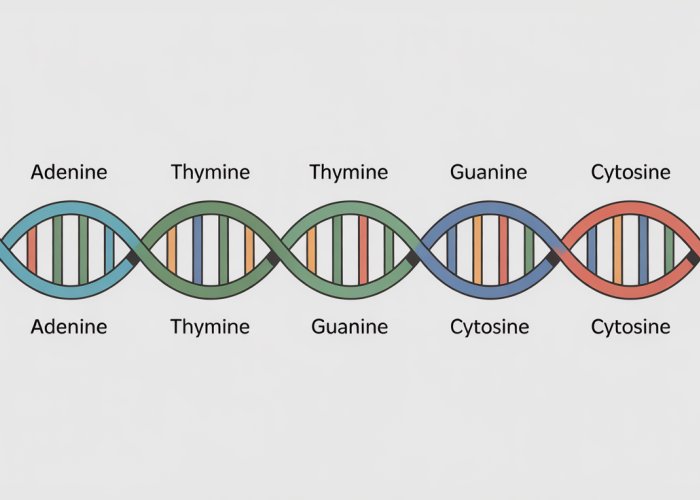
The A-T and G-C Partnerships
The magic of base pairing lies in its specificity. Adenine (A) always pairs with Thymine (T), forming two hydrogen bonds.
Guanine (G) always pairs with Cytosine (C), linked by three hydrogen bonds.
These pairings are not random; they are dictated by the molecular structure of the bases themselves.
The arrangement of hydrogen bond donors and acceptors allows for optimal interactions only between these specific pairs.
This specificity is what ensures the accurate transfer of genetic information from one generation to the next.
Chargaff’s Rules: Unveiling the Quantitative Relationships
Prior to the elucidation of the double helix structure, biochemist Erwin Chargaff observed a crucial pattern in the composition of DNA.
He noted that the amount of Adenine (A) was always approximately equal to the amount of Thymine (T), and the amount of Guanine (G) was always approximately equal to the amount of Cytosine (C).
These observations, now known as Chargaff’s rules, provided a critical clue to Watson and Crick as they pieced together the structure of DNA.
Chargaff’s rules confirmed that there’s a direct, quantitative relationship reflecting A-T and G-C pairings.
These rules strongly supported the hypothesis that these bases are specifically linked.
Base Pairing in DNA Replication
During DNA replication, the double helix unwinds, and each strand serves as a template for the synthesis of a new complementary strand.
The enzyme DNA polymerase uses the principle of base pairing to ensure that the new strand is an exact copy of the original.
As it moves along the template strand, it adds nucleotides that are complementary to the existing bases: A to T, and G to C.
This process ensures the fidelity of DNA replication, minimizing errors and preserving the integrity of the genetic code. Without accurate base pairing, the entire process of DNA replication would collapse, leading to genetic chaos.
Base Pairing in Transcription
Transcription, the process by which RNA is synthesized from a DNA template, also relies heavily on base pairing.
RNA polymerase reads the DNA sequence and synthesizes a complementary RNA molecule, substituting Uracil (U) for Thymine (T).
Thus, Adenine (A) on the DNA template pairs with Uracil (U) on the RNA transcript, while Guanine (G) still pairs with Cytosine (C).
This process allows the genetic information encoded in DNA to be transcribed into RNA, which can then be used to direct protein synthesis. This is the central process in the flow of genetic information.
In essence, base pairing is more than just a structural feature of DNA; it is the driving force behind the processes that sustain life. The seemingly simple rule of A-T and G-C pairing provides the foundation for genetic stability, replication, and expression, ensuring the accurate transmission of hereditary information across generations. Its elegant simplicity belies its profound importance.
Unlocking the Code: Significance of Nucleotide Chain Structure
The unraveling of the nucleotide chain structure was more than just a scientific breakthrough; it was the key that unlocked the very code of life. Understanding how these building blocks assemble provides the foundation for manipulating and interpreting the genetic information that governs all living organisms. This knowledge permeates every facet of modern biology, from decoding individual genomes to engineering novel therapies.
DNA Sequencing: Reading the Book of Life
The ability to determine the precise sequence of nucleotides within a DNA molecule, known as DNA sequencing, has revolutionized our understanding of genetics. Knowing the order of A, T, G, and C bases allows us to "read" the genetic code.
This powerful tool enables scientists to identify genes, understand their function, and pinpoint mutations that contribute to disease.
Techniques like Sanger sequencing and next-generation sequencing rely directly on the principles of nucleotide chain structure to accurately determine the order of bases.
The implications are vast, ranging from personalized medicine to tracing the evolutionary history of species.
The Genome: A Complete Genetic Blueprint
The genome represents the entirety of an organism’s hereditary information, encoded within its DNA. Understanding the nucleotide chain structure is paramount to comprehending the organization and function of the genome.
It allows us to identify genes, regulatory elements, and other important features within the vast expanse of genomic DNA.
Comparative genomics, where the genomes of different species are compared, provides insights into evolutionary relationships and the genetic basis of adaptation.
Analyzing the human genome has yielded invaluable information about human health, disease, and ancestry, paving the way for new diagnostic and therapeutic strategies.
The Central Dogma: From DNA to Protein
The central dogma of molecular biology describes the flow of genetic information from DNA to RNA to protein. This fundamental principle is intricately linked to the structure of the nucleotide chain.
The sequence of nucleotides in DNA dictates the sequence of nucleotides in RNA, which in turn determines the sequence of amino acids in a protein.
This chain of events underlies all biological processes, from metabolism to development.
Understanding the nucleotide chain structure provides the framework for deciphering how genetic information is transcribed into RNA and translated into proteins.
The Role of Polymerases
Polymerases are enzymes that play a crucial role in both DNA replication and RNA transcription. These molecular machines synthesize new nucleotide chains using existing DNA or RNA as a template.
They function by recognizing the specific structure of nucleotides and linking them together in a precise sequence, following the rules of base pairing.
Without polymerases, the faithful duplication of DNA and the synthesis of RNA would be impossible, effectively halting the processes of cell division and gene expression.
Impact on Genetics, Medicine, and Biotechnology
The knowledge of nucleotide chain structure has far-reaching implications across multiple scientific disciplines.
In genetics, it enables the study of inheritance, mutations, and gene regulation.
In medicine, it has led to the development of diagnostic tests, gene therapies, and personalized treatments.
In biotechnology, it facilitates the engineering of organisms with novel traits for applications in agriculture, industry, and environmental remediation.
The ability to manipulate and understand DNA at the molecular level is driving innovation in countless fields, promising further advances in our understanding of life and our ability to improve human health and well-being.
Nucleotide Chain Structure: FAQs
This section answers frequently asked questions about the structure of nucleotide chains in DNA.
What are the key components of a nucleotide?
A nucleotide, the building block of DNA, consists of three parts: a deoxyribose sugar, a phosphate group, and a nitrogenous base (adenine, guanine, cytosine, or thymine). These components link together to form a single nucleotide.
How are nucleotides linked together in a DNA strand?
Nucleotides are joined together in a chain through phosphodiester bonds. These bonds form between the phosphate group of one nucleotide and the sugar of the next. This linkage creates the sugar-phosphate backbone of the nucleotide chain structure.
What determines the sequence of a DNA strand?
The sequence of a DNA strand is determined by the order of the nitrogenous bases. The sequence specifies the genetic information encoded within the DNA molecule. Different sequences of bases lead to different genetic codes.
How does the double helix relate to the nucleotide chain structure?
Two nucleotide chains twist around each other to form the DNA double helix. The sugar-phosphate backbones are on the outside, while the nitrogenous bases pair up (A with T, and G with C) on the inside. This pairing stabilizes the nucleotide chain structure in the double helix formation.
So, there you have it – a peek into the fascinating world of nucleotide chain structure! Hopefully, this gave you a better grasp of how it all works. Keep exploring, and who knows what secrets you’ll uncover next?