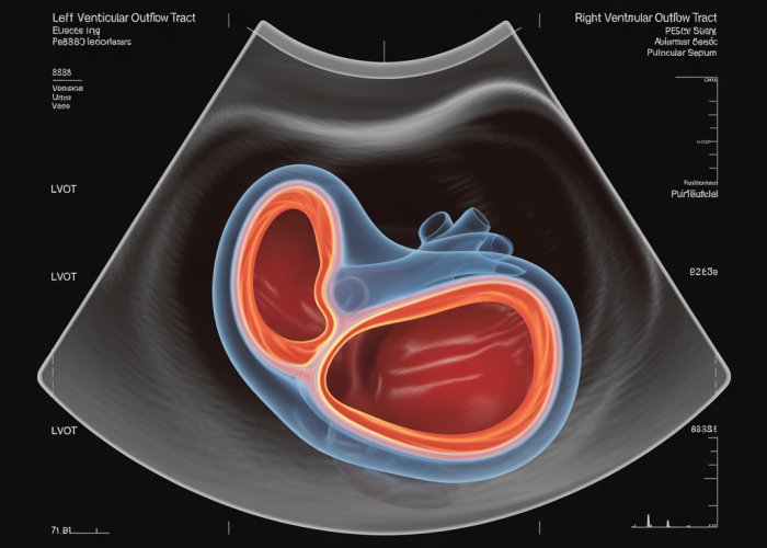
Cardiac function assessment stands as a cornerstone in cardiovascular medicine, and lvot rvot ultrasound plays a vital role. The Left Ventricular Outflow Tract (LVOT), a crucial component, allows blood to flow from the heart to the aorta. Conversely, the Right Ventricular Outflow Tract (RVOT) facilitates blood flow from the heart to the pulmonary artery. These measurements are often interpreted with the knowledge of ASE guidelines (American Society of Echocardiography), the leading organization that provides standards and recommendations on optimal scanning parameters and measurement techniques to further understand the crucial information that lvot rvot ultrasound provides. Therefore, interpreting the data obtained through lvot rvot ultrasound requires a comprehensive understanding of cardiac anatomy and hemodynamics.

The landscape of cardiac diagnostics is constantly evolving, with advancements in imaging technologies playing a pivotal role in improving patient outcomes. Among these advancements, Left Ventricular Outflow Tract (LVOT) and Right Ventricular Outflow Tract (RVOT) ultrasound have emerged as invaluable tools.
These techniques provide critical insights into cardiac function and structure. This has led to their increased use in clinical practice.
The Rising Prominence of LVOT/RVOT Ultrasound
The utilization of LVOT and RVOT ultrasound in cardiac diagnostics has seen a notable surge in recent years. This is due to several factors.
Primarily, the non-invasive nature of echocardiography makes it an attractive option for both initial screening and serial monitoring of cardiac conditions. Unlike more invasive procedures, ultrasound poses minimal risk to the patient, allowing for repeated assessments without significant discomfort or complications.
Furthermore, advancements in ultrasound technology have significantly enhanced image quality and diagnostic capabilities. Modern echocardiography systems offer superior resolution and sophisticated Doppler modalities. This enables clinicians to visualize and quantify blood flow dynamics within the LVOT and RVOT with greater precision.
The ability to accurately assess these outflow tracts is crucial in diagnosing and managing a wide range of cardiovascular diseases.
The Significance of LVOT and RVOT Assessment via Echocardiography
Understanding the intricacies of LVOT and RVOT assessment via echocardiography is paramount for healthcare professionals involved in cardiac care. The LVOT and RVOT are critical components of the heart. They play a central role in systemic and pulmonary circulation, respectively.
Comprehensive evaluation of these outflow tracts provides valuable information about:
- Cardiac function
- Hemodynamic parameters
- The presence of structural abnormalities
This information is essential for accurate diagnosis, risk stratification, and treatment planning in patients with various cardiac conditions.
Echocardiography allows clinicians to visualize the anatomy of the LVOT and RVOT. This includes identifying any obstructions, stenosis, or other structural anomalies.
Doppler ultrasound, a key component of echocardiographic assessment, enables the measurement of blood flow velocities within these outflow tracts.
These measurements are vital for calculating pressure gradients and assessing the severity of any obstruction or stenosis. The information derived from LVOT and RVOT ultrasound can significantly impact clinical decision-making. It also informs the selection of appropriate therapeutic strategies.
Aim and Scope
This article aims to provide healthcare professionals and informed patients with essential knowledge about LVOT and RVOT ultrasound. We strive to offer a comprehensive overview of the underlying principles, techniques, and clinical applications of this important diagnostic modality.
Whether you are a cardiologist, sonographer, medical student, or simply an individual seeking to better understand cardiac health, this resource is designed to enhance your understanding of LVOT and RVOT ultrasound.
By exploring the anatomical and physiological aspects, delving into the echocardiographic techniques used for assessment, and examining the clinical scenarios where LVOT and RVOT ultrasound plays a crucial role. We hope to empower you with the knowledge needed to appreciate the value of this diagnostic tool.
The rising prominence of LVOT and RVOT ultrasound underscores its increasing importance in cardiac diagnostics. To truly appreciate the insights gained from these ultrasound techniques, it’s crucial to first establish a solid understanding of the anatomical and physiological context. This will allow for a better understanding of the blood flow dynamics within these vital cardiac structures.
Anatomy and Physiology: Exploring the LVOT and RVOT
The Left Ventricular Outflow Tract (LVOT) and Right Ventricular Outflow Tract (RVOT) are essential components of the heart, each playing a distinct and critical role in circulatory function. Understanding their anatomical location and physiological function is fundamental to interpreting ultrasound assessments and diagnosing cardiac conditions.
The Left Ventricular Outflow Tract (LVOT)
The LVOT is the conduit through which oxygenated blood exits the left ventricle and enters the aorta, initiating systemic circulation.
Definition and Anatomical Location
The LVOT is not a fixed anatomical structure but rather a dynamic region within the left ventricle. It extends from the mitral valve leaflet to the aortic valve.
It is bordered by the interventricular septum and the anterior mitral valve leaflet. This region is crucial because its size and shape can significantly impact blood flow dynamics.
Role in Systemic Circulation
The LVOT’s primary function is to facilitate the efficient ejection of blood into the aorta. This ensures oxygenated blood reaches all tissues and organs throughout the body.
Any obstruction or abnormality within the LVOT can impede blood flow. This then leads to a decrease in cardiac output and potentially causing symptoms such as shortness of breath, chest pain, and fatigue. Conditions like aortic stenosis or hypertrophic cardiomyopathy directly affect LVOT function.
The Right Ventricular Outflow Tract (RVOT)
The RVOT serves as the pathway for deoxygenated blood to flow from the right ventricle to the pulmonary artery, initiating pulmonary circulation.
Definition and Anatomical Location
The RVOT is located in the superior aspect of the right ventricle. It leads to the pulmonary valve, which then opens into the pulmonary artery.
Unlike the LVOT, the RVOT is more muscular and has a more complex geometry. This makes it susceptible to different types of obstructions and abnormalities.
Role in Pulmonary Circulation
The RVOT’s function is to propel deoxygenated blood to the lungs. There, it undergoes gas exchange, releasing carbon dioxide and absorbing oxygen.
Impairment of RVOT function can lead to pulmonary hypertension, right ventricular hypertrophy, and ultimately, right heart failure. Conditions like pulmonary stenosis and pulmonary hypertension directly impact RVOT function.
Cardiac Physiology Related to Outflow Tracts
The efficient function of both the LVOT and RVOT is dependent on complex interplay of factors. This includes ventricular contractility, valve function, and vascular resistance.
Ventricular contractility directly impacts the force and volume of blood ejected through the outflow tracts.
Valve function ensures unidirectional blood flow, preventing backflow and maintaining efficient circulation.
Vascular resistance in the systemic and pulmonary circulations affects the afterload on the left and right ventricles, respectively.
Understanding the anatomy and physiology of the LVOT and RVOT is crucial for interpreting echocardiographic findings and managing various cardiovascular diseases. Ultrasound assessment of these outflow tracts provides valuable insights into cardiac function. This helps clinicians make informed decisions about diagnosis and treatment.
Echocardiography Essentials: Ultrasound in LVOT/RVOT Assessment
Having established the anatomical and physiological framework of the LVOT and RVOT, it’s time to explore the primary tool used to evaluate these critical cardiac structures: echocardiography. This section delves into the fundamental principles of ultrasound technology, emphasizing its specific application in imaging the heart, particularly the LVOT and RVOT. We’ll also explore the critical role of Doppler techniques in assessing blood flow dynamics and conclude with a brief overview of how cardiologists utilize these tools in clinical practice.
The Foundation: Ultrasound Principles in Cardiac Imaging
Echocardiography, at its core, employs ultrasound waves to create real-time images of the heart. These high-frequency sound waves are emitted by a transducer, which is placed on the patient’s chest.
As the ultrasound waves travel through the body, they encounter different tissues and structures.
A portion of these waves is reflected back to the transducer, providing information about the density, location, and movement of these structures.
This information is then processed by the echocardiography machine to generate a visual representation of the heart, allowing clinicians to assess its size, shape, and function. The ability to visualize the heart in motion is a key advantage of echocardiography.
Doppler Ultrasound: Unveiling Blood Flow Dynamics
While conventional echocardiography provides anatomical information, Doppler ultrasound adds another dimension by assessing blood flow velocity and direction.
This technique leverages the Doppler effect, the change in frequency of a wave (in this case, ultrasound) when it encounters a moving object (red blood cells).
By analyzing the shift in frequency of the reflected ultrasound waves, the echocardiography machine can determine the speed and direction of blood flow within the heart chambers and vessels.
This is particularly crucial in evaluating the LVOT and RVOT, where abnormalities in blood flow can indicate stenosis, regurgitation, or other cardiac conditions.
Color Doppler: Visualizing Flow Patterns
Color Doppler imaging enhances the visualization of blood flow by assigning different colors to blood moving towards or away from the transducer.
Typically, blood flowing towards the transducer is displayed in red, while blood flowing away is displayed in blue.
Variations in color intensity can also indicate the velocity of blood flow, with brighter colors representing higher velocities. Color Doppler provides a quick and intuitive way to assess blood flow patterns within the LVOT and RVOT.
For instance, turbulent flow, often seen in areas of stenosis or regurgitation, is displayed as a mosaic of colors, indicating irregular and chaotic blood movement.
The Cardiologist’s Perspective: Integrating Echocardiography into Clinical Practice
Cardiologists utilize echocardiography as a primary diagnostic tool for a wide range of cardiac conditions. In the context of the LVOT and RVOT, echocardiography helps in:
- Identifying structural abnormalities: such as valve stenosis or hypertrophic cardiomyopathy.
- Assessing the severity of valve disease: by measuring blood flow velocities and pressure gradients.
- Evaluating the impact of cardiac conditions: on overall heart function.
- Guiding treatment decisions: such as the need for medication, intervention, or surgery.
By carefully analyzing the echocardiographic images and Doppler data, cardiologists can gain valuable insights into the health and function of the heart, leading to more accurate diagnoses and more effective treatment plans.
Clinical Applications: Unveiling Pathology with LVOT/RVOT Ultrasound
Having explored the mechanics of ultrasound and its application to visualizing blood flow, we now turn our attention to the clinical utility of LVOT and RVOT echocardiography. This technology is invaluable in diagnosing and monitoring a range of cardiovascular conditions that affect these critical outflow tracts.
Aortic Stenosis: Quantifying the Severity
Aortic stenosis (AS), a narrowing of the aortic valve, is a common and potentially life-threatening condition. LVOT ultrasound plays a pivotal role in assessing its severity.
By measuring the velocity of blood flow through the aortic valve, echocardiography can determine the degree of obstruction.
The Bernoulli Equation and Pressure Gradient Estimation
The Bernoulli equation is applied to estimate the pressure gradient across the stenotic valve. This calculation provides a quantitative measure of the obstruction’s impact on cardiac function.
A higher pressure gradient indicates more severe stenosis, impacting treatment decisions such as valve replacement.
Pulmonary Stenosis: Assessing Right Ventricular Burden
Pulmonary stenosis (PS), a narrowing of the pulmonary valve, obstructs blood flow from the right ventricle to the pulmonary artery.
RVOT ultrasound is crucial in evaluating the severity of PS and its impact on right ventricular function.
Doppler Ultrasound and Peak Velocity
Doppler ultrasound is used to measure the peak velocity of blood flow across the pulmonary valve.
Elevated peak velocities indicate a greater degree of stenosis. This information is used to guide decisions regarding intervention, such as balloon valvuloplasty.
Hypertrophic Cardiomyopathy (HCM): Identifying LVOT Obstruction
Hypertrophic cardiomyopathy (HCM) is a genetic heart condition characterized by thickening of the heart muscle.
This thickening can sometimes lead to LVOT obstruction, particularly during exercise.
Evaluating LVOT Obstruction in HCM
Echocardiography is essential for identifying and quantifying LVOT obstruction in HCM.
Provocative maneuvers, such as exercise or Valsalva, can be used during the ultrasound examination to assess the dynamic nature of the obstruction.
Pulmonary Hypertension: Recognizing RVOT Ultrasound Clues
Pulmonary hypertension (PH) is a condition characterized by elevated blood pressure in the pulmonary arteries.
RVOT ultrasound findings can provide important clues to the presence and severity of PH.
RVOT Findings Indicative of Pulmonary Hypertension
Signs such as right ventricular enlargement, tricuspid regurgitation, and a flattened interventricular septum can suggest PH.
Additionally, pulmonary artery systolic pressure (PASP) can be estimated using Doppler echocardiography.
Right Ventricular Outflow Tract Obstruction (RVOTO)
RVOTO encompasses a variety of conditions that impede blood flow from the right ventricle.
Causes and Ultrasound Findings
Potential causes include congenital abnormalities, tumors, and thrombi. Ultrasound can help visualize the obstruction and assess its impact on right ventricular function.
Characteristic ultrasound findings may include RV dilation, paradoxical septal motion, and elevated RVOT velocities.
Left Ventricular Outflow Tract Obstruction (LVOTO)
LVOTO refers to any obstruction in the path of blood flow leaving the left ventricle, excluding aortic valve stenosis.
Causes and Ultrasound Findings
Causes of LVOTO can include subaortic membranes, hypertrophic cardiomyopathy, and mitral valve abnormalities.
Echocardiography helps visualize the obstructing lesion, assess its severity, and evaluate left ventricular function. Doppler studies are crucial for quantifying the pressure gradient across the obstruction.
Examination and Interpretation: LVOT/RVOT Ultrasound in Practice
Having explored the clinical scenarios where LVOT and RVOT ultrasound proves invaluable, it’s essential to understand how this diagnostic tool is employed in real-world settings. Let’s delve into the practical aspects of the examination process and the critical role of interpretation in guiding patient care.
The LVOT/RVOT Ultrasound Examination: A Concise Overview
The LVOT/RVOT ultrasound examination is a non-invasive procedure that utilizes sound waves to create detailed images of the heart’s outflow tracts.
The examination typically involves placing a transducer on the patient’s chest.
The transducer emits ultrasound waves that bounce off the heart’s structures, providing real-time visualization of the LVOT and RVOT.
Various imaging modalities, such as 2D imaging, Doppler ultrasound, and color Doppler, are employed to assess the anatomy and function of these outflow tracts.
The Crucial Role of the Echocardiography Technician (Sonographer)
The Echocardiography Technician, also known as a Sonographer, is a highly skilled healthcare professional who plays a pivotal role in the LVOT/RVOT ultrasound examination.
The Sonographer is responsible for acquiring high-quality images of the heart, ensuring optimal visualization of the LVOT and RVOT.
They must possess a thorough understanding of cardiac anatomy, physiology, and pathology to accurately capture relevant data.
The Sonographer’s expertise in manipulating the ultrasound equipment and adjusting imaging parameters is critical for obtaining diagnostic-quality images.
They work closely with the Cardiologist, providing valuable insights and preliminary findings that aid in the interpretation process.
Key Measurements and Parameters: Unveiling Cardiac Function
LVOT/RVOT ultrasound provides a wealth of information about cardiac function, enabling clinicians to assess the severity of various cardiovascular conditions.
Several key measurements and parameters are routinely assessed during the examination.
Cardiac Output, a measure of the volume of blood pumped by the heart per minute, is an important indicator of overall cardiac function.
Stroke Volume, the amount of blood ejected by the left ventricle with each heartbeat, is another essential parameter.
These measurements, along with assessments of blood flow velocity, pressure gradients, and valve function, provide a comprehensive evaluation of LVOT and RVOT performance.
Accurate Interpretation: The Cornerstone of Diagnosis and Management
The accurate interpretation of LVOT/RVOT ultrasound findings is paramount for making informed diagnoses and guiding appropriate treatment strategies.
Cardiologists meticulously analyze the images and measurements obtained during the examination, correlating them with the patient’s clinical history and other diagnostic data.
Misinterpretation of ultrasound findings can have significant consequences, leading to delayed or inappropriate treatment decisions.
For example, accurately quantifying the severity of aortic stenosis through LVOT ultrasound is crucial for determining the optimal timing for valve replacement surgery.
Similarly, RVOT ultrasound findings can help differentiate between various causes of pulmonary hypertension, guiding targeted therapies.
Therefore, a collaborative approach involving skilled Sonographers and experienced Cardiologists is essential for ensuring accurate interpretation and optimal patient management.
FAQs: LVOT RVOT Ultrasound
Here are some frequently asked questions about LVOT RVOT ultrasounds to help you better understand the procedure and its purpose.
What exactly does LVOT RVOT stand for?
LVOT stands for Left Ventricular Outflow Tract, and RVOT stands for Right Ventricular Outflow Tract. These are the areas where blood exits the left and right ventricles of your heart, respectively. A lvot rvot ultrasound is a specialized imaging test that focuses on evaluating the function of these areas.
Why would I need a lvot rvot ultrasound?
This type of ultrasound is often ordered to assess for obstructions or abnormalities in the outflow tracts. This could include conditions like stenosis (narrowing) or other structural issues impacting blood flow. It helps doctors diagnose and manage certain heart conditions.
What happens during a lvot rvot ultrasound?
The process is similar to a standard echocardiogram. A technician applies gel to your chest and uses a transducer to capture images of your heart. The ultrasound waves allow visualization of the LVOT and RVOT. It is a non-invasive and painless procedure.
How is an lvot rvot ultrasound different from a regular echocardiogram?
While a standard echocardiogram provides a comprehensive overview of the heart, an lvot rvot ultrasound focuses specifically on detailed examination of the left and right ventricular outflow tracts. This allows for a more precise assessment of these areas and any related conditions.
Alright, that pretty much covers the essentials of lvot rvot ultrasound! Hope this helped clear things up. Now you’ve got the basics, so go ahead and explore, keep learning, and see what else you can discover about this super-useful diagnostic tool.