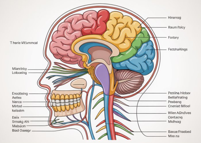
Understanding the intricate network of the human nervous system hinges on a firm grasp of the cranial nerves. Neuroanatomy, the study of the nervous system’s structure, provides the essential framework for identifying and understanding these vital components. Effective medical education often employs detailed diagrams and models to help students accurately label cranial nerves. A comprehensive knowledge of these twelve pairs of nerves, responsible for a multitude of sensory and motor functions, is absolutely critical for professionals within the healthcare industry. With a detailed visual guide you will be able to easily label cranial nerves.
The human nervous system, a vast and complex network, governs everything from our simplest reflexes to our most profound thoughts. At the heart of this system lies a specialized set of nerves known as the cranial nerves.
These twelve pairs of nerves emerge directly from the brain and brainstem, serving as vital communication lines between the brain and various structures in the head, neck, and torso.
Understanding their anatomy and function is paramount, not only for students of medicine and related fields, but also for any professional involved in the diagnosis and treatment of neurological conditions.
The Cranial Nerves Defined: Gateways to Sensory and Motor Function
Cranial nerves are distinct from spinal nerves, which originate from the spinal cord.
Each cranial nerve is designated by a Roman numeral (I-XII) and a name that reflects its primary function or anatomical course.
They play an indispensable role in a diverse range of functions, including:
- Sensory perception (sight, smell, taste, hearing, and touch).
- Motor control of facial muscles, eye movement, tongue movement, swallowing, and head/shoulder movement.
- Parasympathetic regulation of vital functions such as heart rate, digestion, and glandular secretions.
Without these nerves, we would be unable to experience the world around us or effectively interact with it.
The Critical Importance of Accurate Labeling and Understanding
The intricacies of cranial nerve anatomy demand precision.
Accurate labeling is essential for several reasons:
- Effective Communication: Standardized labeling allows healthcare professionals to communicate clearly and concisely about specific nerves and their associated structures, facilitating accurate diagnosis and treatment planning.
- Diagnostic Precision: Identifying the precise location of a nerve lesion is crucial for determining the underlying cause of neurological deficits.
- Surgical Accuracy: During neurosurgical procedures, a detailed understanding of cranial nerve anatomy is paramount to avoid iatrogenic injury and preserve neurological function.
- Educational Foundation: For students, mastering the accurate labeling and anatomical pathways of the cranial nerves lays the foundation for future clinical practice.
Misidentification or misunderstanding can lead to serious consequences, underscoring the need for comprehensive and precise anatomical knowledge.
A Comprehensive, Visually-Driven Approach
This guide champions a visually-driven approach to understanding and labeling the cranial nerves.
The power of visual learning cannot be overstated, particularly when grappling with complex anatomical structures.
By combining clear, concise explanations with detailed illustrations, diagrams, and potentially even 3D models, this method aims to:
- Enhance comprehension of cranial nerve anatomy and function.
- Facilitate accurate labeling of key structures.
- Improve retention of information.
- Provide a valuable resource for students, educators, and practicing professionals.
The goal is to provide a resource that simplifies a traditionally challenging subject, empowering readers with the confidence and knowledge to navigate the intricate world of cranial nerves.
The Foundation: Anatomy of the Brain and Brainstem
Before delving into the specifics of each cranial nerve, it’s critical to establish a solid understanding of the anatomical structures that serve as their origin and processing centers. These include the brain itself and, most crucially, the brainstem. Together, these structures form the central command hub for cranial nerve function.
The Brain’s Supervisory Role in Cranial Nerve Function
While the brainstem serves as the primary origin point for most cranial nerves, the brain, particularly the cerebral cortex, plays a vital supervisory role. Sensory information gathered by cranial nerves such as the olfactory (CN I), optic (CN II), and vestibulocochlear (CN VIII) nerves is ultimately processed and interpreted within specific cortical regions.
For instance, visual information travels from the optic nerve to the visual cortex in the occipital lobe, allowing us to perceive and understand what we see. Similarly, auditory information from the vestibulocochlear nerve is processed in the auditory cortex of the temporal lobe, enabling us to hear and interpret sounds.
Furthermore, the brain exerts higher-level control over motor functions regulated by cranial nerves. The motor cortex in the frontal lobe initiates voluntary movements, which are then relayed to cranial nerve motor nuclei in the brainstem to control muscles of the face, head, and neck.
This intricate interplay between the brain and brainstem underscores the importance of understanding both structures to fully grasp cranial nerve function.
The Brainstem: Origin Point and Relay Station
The brainstem, comprised of the midbrain, pons, and medulla oblongata, is the primary origin point for all but two cranial nerves (CN I and CN II). It serves as a critical relay station, connecting the brain to the spinal cord and housing the nuclei of cranial nerves III through XII.
Within the brainstem, specific nuclei act as the functional centers for each cranial nerve. These nuclei contain the cell bodies of the neurons that form the cranial nerves, and they receive input from various brain regions, integrating this information to control specific functions.
Key brainstem structures that are particularly relevant to cranial nerve function include:
-
Midbrain: Contains nuclei for the oculomotor (CN III) and trochlear (CN IV) nerves, which control eye movement.
-
Pons: Houses nuclei for the trigeminal (CN V), abducens (CN VI), and facial (CN VII) nerves, involved in facial sensation, eye movement, facial expression, and taste.
-
Medulla Oblongata: Contains nuclei for the vestibulocochlear (CN VIII), glossopharyngeal (CN IX), vagus (CN X), accessory (CN XI), and hypoglossal (CN XII) nerves, responsible for hearing, balance, taste, swallowing, parasympathetic control, and head/shoulder/tongue movement.
Cranial Nerve Pathways: The Routes of Communication
Understanding the pathways of cranial nerves is essential for comprehending how they transmit sensory and motor information between the brain and the periphery. These pathways can be complex, involving multiple synapses and crossing over to the opposite side of the brain.
Sensory pathways typically involve a series of neurons that relay information from sensory receptors to the brainstem nuclei and ultimately to the cerebral cortex. For example, the pathway for facial sensation (trigeminal nerve) involves three neurons:
- The first neuron carries sensory information from the face to the trigeminal ganglion.
- The second neuron relays the information to the trigeminal nucleus in the brainstem.
- The third neuron projects from the brainstem to the thalamus and then to the sensory cortex.
Motor pathways originate in the motor cortex of the brain and descend to the brainstem, where they synapse with motor neurons in the cranial nerve nuclei. These motor neurons then project to muscles in the head and neck, controlling their movement.
Damage to any point along these pathways can result in specific neurological deficits, highlighting the importance of a thorough understanding of their anatomy. For example, a lesion along the facial nerve pathway can lead to facial paralysis (Bell’s Palsy), while damage to the optic nerve can result in vision loss.
By understanding the brain’s role, the brainstem’s central function, and the pathways of cranial nerves, we establish a framework for a deeper exploration of these essential components of the nervous system.
The brain and brainstem, with their intricate architecture and collaborative functions, set the stage for understanding the cranial nerves. Now, we turn our attention to the individual players in this neurological symphony. Each of the twelve cranial nerves possesses a unique role, a defined pathway, and distinct anatomical landmarks that allow for identification.
A Detailed Look at Each Cranial Nerve (CN I – CN XII)
This section serves as the core of our exploration, providing an in-depth examination of the twelve cranial nerves (CN I-CN XII). For each nerve, we will discuss its primary function, trace its anatomical pathway, and highlight key visual identification points to aid in accurate labeling and understanding.
Olfactory Nerve (CN I): The Sense of Smell
The olfactory nerve (CN I) is responsible for our sense of smell. This sensory nerve transmits information from the nasal cavity directly to the brain, enabling us to perceive and differentiate between various odors.
Function: Detecting Odors
The primary function of the olfactory nerve is olfaction, the process of smelling. Specialized olfactory receptor neurons located in the nasal epithelium detect volatile odor molecules.
These neurons then transmit signals to the olfactory bulb in the brain. This sensory function is vital for detecting potential dangers like smoke or spoiled food and also contributes significantly to our sense of taste.
Pathway: From Nose to Brain
The olfactory pathway begins in the nasal cavity, where olfactory receptor neurons detect odorants. These neurons send axons through the cribriform plate, a sieve-like structure in the ethmoid bone, to reach the olfactory bulb.
From the olfactory bulb, signals travel along the olfactory tract to the olfactory cortex in the temporal lobe of the brain for further processing. Unlike other sensory pathways, the olfactory pathway does not relay through the thalamus before reaching the cortex.
Visual Label: Olfactory Bulb and Tract
The olfactory bulb, located just above the nasal cavity, is a key visual landmark. It appears as an oval-shaped structure where olfactory nerve fibers converge.
The olfactory tract, a bundle of nerve fibers extending from the olfactory bulb, is also visible. These structures serve as important visual identifiers for the olfactory nerve.
Optic Nerve (CN II): The Sense of Sight
The optic nerve (CN II) is dedicated to vision, transmitting visual information from the retina of the eye to the brain. This allows us to perceive shapes, colors, and movement.
Function: Perceiving Visual Information
The optic nerve’s function is to transmit visual information. Light enters the eye and is focused onto the retina, where photoreceptor cells (rods and cones) convert light into electrical signals.
These signals are then relayed to ganglion cells, whose axons converge to form the optic nerve. This sensory nerve is essential for sight and visual perception.
Pathway: From Eye to Brain
The optic nerve originates in the retina of the eye and exits the orbit through the optic canal. The two optic nerves (one from each eye) meet at the optic chiasm, located at the base of the brain.
At the optic chiasm, some fibers from each nerve cross over to the opposite side of the brain. This crossover allows for binocular vision and depth perception.
After the optic chiasm, the nerve fibers continue as the optic tracts to the lateral geniculate nucleus (LGN) of the thalamus. From the LGN, information is relayed to the visual cortex in the occipital lobe for processing.
Visual Label: Optic Chiasm and Optic Tract
The optic chiasm, where the optic nerves partially cross, is a crucial visual landmark. It is located anterior to the pituitary gland.
The optic tracts, which continue from the optic chiasm, are also important visual identifiers as they course towards the thalamus.
Oculomotor Nerve (CN III): Eye Movement and Pupil Control
The oculomotor nerve (CN III) primarily controls eye movement and is also responsible for pupillary constriction and eyelid elevation. This motor nerve innervates most of the eye muscles.
Function: Controlling Eye Muscles
The oculomotor nerve controls four of the six extraocular muscles responsible for eye movement: the superior rectus, inferior rectus, medial rectus, and inferior oblique muscles.
It also controls the levator palpebrae superioris muscle, which elevates the eyelid. Additionally, the oculomotor nerve carries parasympathetic fibers that control pupillary constriction via the sphincter pupillae muscle and accommodation of the lens for near vision.
Pathway: From Brainstem to Eye
The oculomotor nerve originates in the midbrain and exits the brainstem anteriorly. It then passes through the cavernous sinus and enters the orbit through the superior orbital fissure.
Within the orbit, the nerve divides into superior and inferior branches, which innervate the respective eye muscles. The parasympathetic fibers travel with the inferior branch to innervate the pupillary constrictor and ciliary muscles.
Visual Label: Superior Orbital Fissure
The superior orbital fissure, the bony opening through which the oculomotor nerve enters the orbit, is a key visual landmark. It is located in the posterior aspect of the orbit and is a crucial point for identifying the nerve’s entry into the eye socket.
Trochlear Nerve (CN IV): Superior Oblique Muscle Control
The trochlear nerve (CN IV) controls a single muscle: the superior oblique muscle, which is responsible for downward and outward eye movement and torsion (internal rotation) of the eyeball.
Function: Controlling the Superior Oblique Muscle
The trochlear nerve exclusively innervates the superior oblique muscle. This muscle is responsible for depressing, abducting, and internally rotating the eye.
These movements are critical for tasks like looking down and to the side. Dysfunction of this nerve can lead to diplopia (double vision), particularly when looking downward.
Pathway: Unique Dorsal Exit
The trochlear nerve is unique among the cranial nerves. It is the only cranial nerve to exit the brainstem dorsally. It arises from the posterior aspect of the midbrain, wraps around the brainstem, and then travels anteriorly.
Similar to the oculomotor nerve, it passes through the cavernous sinus and enters the orbit through the superior orbital fissure to innervate the superior oblique muscle.
Visual Label: Dorsal Exit from Brainstem
The dorsal exit of the trochlear nerve from the brainstem is a distinctive visual identifier. This characteristic feature distinguishes it from all other cranial nerves, which exit ventrally or laterally.
Trigeminal Nerve (CN V): Facial Sensation and Mastication
The trigeminal nerve (CN V) is a mixed nerve with both sensory and motor functions. It is responsible for providing sensation to the face, oral cavity, and nasal cavity, as well as controlling the muscles of mastication (chewing).
Function: Sensory and Motor Control
The trigeminal nerve has both sensory and motor functions. Its sensory function includes providing tactile, pain, and temperature sensation to the face, forehead, scalp, cornea, nasal cavity, and oral cavity.
Its motor function controls the muscles of mastication (masseter, temporalis, medial pterygoid, and lateral pterygoid muscles), which are essential for chewing. The trigeminal nerve is the largest cranial nerve and plays a critical role in facial sensation and motor function.
Pathway: From Face to Brainstem
The trigeminal nerve originates from the pons region of the brainstem. It has three major branches: the ophthalmic (V1), maxillary (V2), and mandibular (V3) nerves.
The ophthalmic nerve (V1) provides sensation to the forehead, upper eyelid, and cornea. The maxillary nerve (V2) provides sensation to the lower eyelid, cheek, nasal cavity, and upper lip.
The mandibular nerve (V3) provides sensation to the lower lip, chin, and anterior tongue, and also innervates the muscles of mastication. These branches traverse through specific foramina in the skull to reach their respective target areas.
Visual Label: Three Major Branches
The three branches of the trigeminal nerve—ophthalmic (V1), maxillary (V2), and mandibular (V3)—are crucial visual landmarks. Identifying these branches and their distribution patterns aids in understanding the nerve’s complex sensory and motor functions.
Abducens Nerve (CN VI): Lateral Rectus Muscle Control
The abducens nerve (CN VI) controls a single muscle: the lateral rectus muscle, which is responsible for abduction (outward movement) of the eye.
Function: Abducting the Eye
The sole function of the abducens nerve is to innervate the lateral rectus muscle, which abducts the eye. This movement allows us to look laterally (to the side).
Damage to this nerve results in diplopia (double vision) when attempting to look towards the affected side.
Pathway: Long Intracranial Course
The abducens nerve originates from the pons region of the brainstem. It has a relatively long intracranial course, traveling anteriorly along the base of the skull before entering the cavernous sinus.
It then enters the orbit through the superior orbital fissure and innervates the lateral rectus muscle. Its long intracranial course makes it vulnerable to injury from increased intracranial pressure or trauma.
Visual Label: Intracranial Course
The long intracranial course of the abducens nerve is an important visual marker. Tracing its pathway from the brainstem, along the base of the skull, and through the cavernous sinus is key to its identification.
Facial Nerve (CN VII): Facial Expression, Taste, and Salivation
The facial nerve (CN VII) is a mixed nerve responsible for controlling facial expression, taste sensation from the anterior two-thirds of the tongue, and salivation.
Function: Multiple Roles in Sensation and Motor Control
The facial nerve has diverse functions including motor control of facial expression muscles. It is responsible for taste sensation from the anterior two-thirds of the tongue and controls salivation from the submandibular and sublingual glands. It also innervates the stapedius muscle of the middle ear.
Pathway: Complex Pathway Through Skull
The facial nerve originates from the pons region of the brainstem and enters the temporal bone through the internal acoustic meatus. It then travels through the facial canal within the temporal bone, giving off branches to the stapedius muscle and the chorda tympani nerve (which carries taste fibers from the tongue).
The nerve exits the skull through the stylomastoid foramen and enters the parotid gland, where it divides into five major branches that innervate the muscles of facial expression.
Visual Label: Branches within the Parotid Gland
The branches of the facial nerve within the parotid gland are significant visual landmarks. While the nerve itself is deep, understanding its branching pattern in this region is essential for surgical procedures to avoid nerve damage.
Vestibulocochlear Nerve (CN VIII): Hearing and Balance
The vestibulocochlear nerve (CN VIII) is a sensory nerve responsible for hearing and balance. It has two divisions: the cochlear nerve (for hearing) and the vestibular nerve (for balance).
Function: Hearing and Equilibrium
The vestibulocochlear nerve is responsible for both hearing and balance. The cochlear nerve transmits auditory information from the cochlea to the brain, allowing us to perceive sounds.
The vestibular nerve transmits information about head position and movement from the vestibular apparatus to the brain, which is essential for maintaining balance and spatial orientation.
Pathway: From Inner Ear to Brainstem
The vestibulocochlear nerve originates from the inner ear. The cochlear nerve arises from the cochlea and the vestibular nerve arises from the semicircular canals and otolith organs.
Both divisions travel together through the internal acoustic meatus to enter the brainstem at the pontomedullary junction. From there, auditory and vestibular information is relayed to various brain regions for processing.
Visual Label: Cochlear and Vestibular Components
The cochlear and vestibular components of the nerve are important visual identifiers. While the nerve itself is deep, understanding the location of the cochlea and vestibular apparatus within the inner ear is key to understanding the origin of the nerve’s signals.
Glossopharyngeal Nerve (CN IX): Taste, Swallowing, and Salivation
The glossopharyngeal nerve (CN IX) is a mixed nerve involved in taste sensation, swallowing, salivation, and monitoring blood pressure.
Function: Diverse Sensory and Motor Roles
The glossopharyngeal nerve has various functions. It is responsible for taste sensation from the posterior one-third of the tongue.
It controls swallowing via the stylopharyngeus muscle, stimulates salivation from the parotid gland, and monitors blood pressure via the carotid sinus. The nerve also contributes to the gag reflex.
Pathway: From Mouth to Brainstem
The glossopharyngeal nerve originates from the medulla oblongata of the brainstem. It exits the skull through the jugular foramen and travels to the tongue, pharynx, and parotid gland.
Along its course, it gives off branches that provide taste sensation to the posterior tongue, motor innervation to the stylopharyngeus muscle, and parasympathetic innervation to the parotid gland.
Visual Label: Role in the Gag Reflex
The glossopharyngeal nerve’s role in the gag reflex provides a visual marker. Stimulation of the posterior pharynx elicits the gag reflex, which tests the function of the glossopharyngeal nerve (sensory component) and the vagus nerve (motor component).
Vagus Nerve (CN X): Parasympathetic Control and Visceral Sensation
The vagus nerve (CN X) is a mixed nerve with extensive distribution throughout the thorax and abdomen. It plays a critical role in parasympathetic control of visceral organs, taste sensation, and swallowing.
Function: Extensive Parasympathetic Control
The vagus nerve is the primary nerve of the parasympathetic nervous system, controlling heart rate, digestion, and respiratory function. It provides motor innervation to the muscles of the pharynx and larynx, which are important for swallowing and speech.
It also carries taste sensation from the epiglottis and provides sensory innervation to the visceral organs of the thorax and abdomen. The vagus nerve has the most extensive distribution of all cranial nerves.
Pathway: Thorax and Abdomen
The vagus nerve originates from the medulla oblongata of the brainstem and exits the skull through the jugular foramen. It travels down the neck and into the thorax and abdomen, giving off branches to various organs along the way.
It innervates the heart, lungs, esophagus, stomach, intestines, liver, pancreas, and kidneys. Its extensive distribution reflects its widespread parasympathetic control.
Visual Label: Body-wide Distribution
The extensive distribution of the vagus nerve throughout the body is a key visual concept. Understanding the organs innervated by the vagus nerve and its pathway through the thorax and abdomen is essential for comprehending its function.
Accessory Nerve (CN XI): Head and Shoulder Movement
The accessory nerve (CN XI) is a motor nerve that controls the sternocleidomastoid and trapezius muscles, which are responsible for head and shoulder movement.
Function: Controlling Neck Muscles
The accessory nerve controls the sternocleidomastoid and trapezius muscles. The sternocleidomastoid muscle is responsible for head rotation and flexion, while the trapezius muscle is responsible for shoulder elevation and scapular retraction.
These muscles are essential for head and shoulder movement. Weakness of these muscles can result in difficulty turning the head or shrugging the shoulders.
Pathway: Spinal Cord and Brainstem
The accessory nerve has a unique origin. It has both a cranial root, originating from the medulla oblongata, and a spinal root, originating from the upper cervical spinal cord.
The spinal root ascends through the foramen magnum to join the cranial root, and together they exit the skull through the jugular foramen. The nerve then travels to innervate the sternocleidomastoid and trapezius muscles.
Visual Label: Spinal Root
The spinal root of the accessory nerve is a distinctive visual feature. It is the only cranial nerve with a spinal origin. Identifying this root helps distinguish it from other cranial nerves.
Hypoglossal Nerve (CN XII): Tongue Movement
The hypoglossal nerve (CN XII) is a motor nerve that controls the muscles of the tongue, which are essential for speech, swallowing, and manipulating food.
Function: Controlling Tongue Muscles
The hypoglossal nerve innervates all the intrinsic and most of the extrinsic muscles of the tongue. These muscles control tongue movement, which is critical for speech articulation, swallowing, and manipulating food in the mouth.
Weakness of the tongue muscles can result in difficulty speaking, swallowing, or moving food around in the mouth.
Pathway: From Brainstem to Tongue
The hypoglossal nerve originates from the medulla oblongata of the brainstem. It exits the skull through the hypoglossal canal and travels to the tongue.
Along its course, it innervates the various tongue muscles, allowing for precise control of tongue movement.
Visual Label: Hypoglossal Canal
The hypoglossal canal, the bony opening through which the hypoglossal nerve exits the skull, is a key visual landmark. Identifying this canal helps trace the nerve’s pathway from the brainstem to the tongue.
Visual Aids for Effective Learning
The intricate network of cranial nerves, with their diverse functions and complex pathways, can present a significant challenge for students and professionals alike. Navigating this intricate anatomy requires more than just rote memorization. It demands a deep spatial understanding that can be significantly enhanced through the strategic use of visual aids.
Visual aids serve as invaluable tools in demystifying the complexities of cranial nerve anatomy, transforming abstract concepts into tangible and relatable representations. By leveraging the power of visual learning, individuals can more effectively grasp the spatial relationships, pathways, and functions of these crucial neurological structures.
The Power of Visualization in Neuroanatomy
The human brain is inherently wired to process visual information more efficiently than textual data. Visual aids tap into this inherent cognitive advantage, facilitating a deeper and more lasting understanding of complex anatomical concepts.
When studying cranial nerves, a static textual description can often fall short in conveying the three-dimensional relationships and intricate pathways of these structures. Visual aids, however, offer a dynamic and intuitive way to explore these complex relationships, allowing learners to visualize the origins, courses, and terminations of each nerve in a manner that text alone simply cannot replicate.
Recommended Visual Tools
To maximize learning outcomes, it’s crucial to select visual aids that are both accurate and informative. Here are two highly recommended types of visual tools:
-
Labeled Diagrams: Well-labeled diagrams provide a clear and concise visual representation of cranial nerve anatomy. Look for diagrams that highlight key landmarks, such as the origins of the nerves in the brainstem, their pathways through the skull, and their target organs. The labels should be detailed and accurate, providing a comprehensive overview of each nerve’s anatomy.
Effective diagrams should also incorporate color-coding to differentiate between individual nerves, further enhancing visual clarity and facilitating easier identification.
-
3D Models: Three-dimensional models, whether physical or digital, offer an unparalleled level of spatial understanding. These models allow learners to rotate and explore the cranial nerves from multiple perspectives, revealing the intricate relationships between these structures and their surrounding anatomical landmarks.
Digital 3D models often provide interactive features such as the ability to isolate individual nerves, view cross-sections, and access detailed anatomical information. These interactive capabilities can significantly enhance the learning experience, fostering a deeper and more intuitive understanding of cranial nerve anatomy.
Example of an Effective Labeled Diagram
A high-quality, labeled diagram should clearly depict the following features for each cranial nerve:
- Its point of origin within the brain or brainstem.
- Its course through the skull, including any relevant foramina or fissures.
- Its target organs or tissues.
- Any significant branches or subdivisions.
The diagram should also include labels for surrounding anatomical structures, such as the skull bones, brain regions, and major blood vessels. The use of clear, concise labels and color-coding is essential for maximizing the diagram’s effectiveness.
For instance, a diagram illustrating the trigeminal nerve (CN V) should clearly depict its three branches (ophthalmic, maxillary, and mandibular) as well as their respective distributions across the face. Similarly, a diagram of the vagus nerve (CN X) should highlight its extensive distribution throughout the thorax and abdomen, illustrating its role in parasympathetic control.
By incorporating such visual aids into their study routine, students and professionals can significantly enhance their understanding of cranial nerve anatomy, leading to improved diagnostic and clinical skills.
Visual aids are powerful tools in learning, offering a concrete way to understand abstract concepts. But beyond seeing, how can one truly remember the intricacies of the cranial nerves? This is where the art of memorization steps in, employing clever techniques to transform a list of names and functions into lasting knowledge.
Mnemonic Devices and Learning Strategies
Mnemonic devices and strategic learning techniques are indispensable tools for mastering the cranial nerves. These methods leverage the brain’s natural inclination for patterns and associations, transforming seemingly arbitrary information into memorable nuggets of knowledge. By employing these strategies, both students and seasoned professionals can solidify their understanding and recall of these essential neurological components.
The Power of Mnemonics: Bridging Memory Gaps
Mnemonic devices are memory aids that use vivid associations, rhymes, or acronyms to encode information in a more accessible format. They are particularly useful when dealing with lists or sequences, such as the names and order of the cranial nerves.
The effectiveness of mnemonics stems from their ability to engage multiple cognitive processes. By linking new information to existing knowledge or creating memorable visual images, these devices enhance encoding and retrieval. This makes them a powerful tool for long-term retention.
Decoding the Cranial Nerves: Examples of Effective Mnemonics
Several popular mnemonics exist to aid in remembering the names and order of the cranial nerves. Here are a few examples:
-
"Oh Oh Oh To Touch And Feel Very Good Velvet AH": This classic mnemonic represents the first letter of each cranial nerve: Olfactory, Optic, Oculomotor, Trochlear, Trigeminal, Abducens, Facial, Vestibulocochlear, Glossopharyngeal, Vagus, Accessory, Hypoglossal.
-
"On Old Olympus’ Towering Top A Finn And German Viewed Some Hops": Another variation for remembering the names.
-
"Some Say Marry Money But My Brother Says Big Brains Matter More": This mnemonic helps recall whether each nerve is Sensory (S), Motor (M), or Both (B) in function.
Choosing a mnemonic that resonates personally is key to its effectiveness. Some learners prefer visual mnemonics, creating mental images associated with each nerve. Others find auditory mnemonics, such as rhymes or songs, more helpful.
Beyond Mnemonics: Effective Study Techniques
While mnemonics are valuable, a comprehensive approach to learning the cranial nerves should include other effective study techniques:
-
Spaced Repetition: Reviewing material at increasing intervals strengthens memory over time. This technique combats the forgetting curve, ensuring long-term retention.
-
Active Recall: Testing yourself regularly, without referring to notes, forces your brain to retrieve information, solidifying learning. Flashcards and practice questions are excellent tools for active recall.
-
Teach Someone Else: Explaining the cranial nerves to another person reinforces your own understanding and identifies areas where you may need further clarification.
-
Concept Mapping: Creating visual diagrams that connect the cranial nerves to their functions, pathways, and associated structures can enhance understanding and retention.
-
Clinical Correlation: Connecting the anatomy and function of each nerve to real-world clinical scenarios makes the information more relevant and memorable. Consider, for example, how damage to the facial nerve (CN VII) can result in Bell’s palsy, causing facial paralysis.
-
Integrate multiple resources: Leverage textbooks, online resources, and anatomical models to create a multi-sensory learning experience. The use of a variety of media helps consolidate understanding.
Tailoring Strategies for Individual Learning Styles
Not all learning strategies are created equal. It is crucial to tailor your approach to your individual learning style.
-
Visual learners benefit from diagrams, illustrations, and videos.
-
Auditory learners thrive on lectures, discussions, and mnemonic songs.
-
Kinesthetic learners learn best through hands-on activities, such as dissecting models or drawing diagrams.
By identifying your preferred learning style and adapting your study techniques accordingly, you can optimize your learning and retention of the cranial nerves.
Visual aids are powerful tools in learning, offering a concrete way to understand abstract concepts. But beyond seeing, how can one truly remember the intricacies of the cranial nerves? This is where the art of memorization steps in, employing clever techniques to transform a list of names and functions into lasting knowledge.
Clinical Significance of Cranial Nerves: Linking Anatomy to Patient Care
The true value of mastering cranial nerve anatomy lies not just in academic achievement, but in its profound clinical significance. These nerves are the direct interface between the brain and critical sensory and motor functions of the head and neck. Understanding their pathways and functions is paramount for accurate diagnosis and effective treatment of a wide range of neurological disorders.
Cranial Nerve Dysfunction: A Window into Neurological Health
Dysfunction of one or more cranial nerves can be an early indicator of various underlying conditions. These conditions range from localized nerve damage to widespread systemic diseases. The specific pattern of deficits observed during a cranial nerve examination provides valuable clues. These clues assist clinicians in pinpointing the location and nature of the underlying pathology.
For example, a patient presenting with sudden onset facial drooping and an inability to close one eye might be suffering from Bell’s Palsy, a condition affecting the facial nerve (CN VII). Conversely, a patient experiencing intense, stabbing facial pain triggered by simple actions like chewing or shaving could be diagnosed with Trigeminal Neuralgia, a disorder involving the trigeminal nerve (CN V).
Common Cranial Nerve Disorders: An Overview
Several disorders commonly affect the cranial nerves, each with distinct characteristics and implications:
-
Bell’s Palsy: As mentioned, this involves inflammation or compression of the facial nerve (CN VII). It results in unilateral facial paralysis.
-
Trigeminal Neuralgia: Characterized by excruciating pain along the distribution of the trigeminal nerve (CN V), often triggered by minimal stimulation.
-
Optic Neuritis: Inflammation of the optic nerve (CN II). It can cause sudden vision loss and is often associated with multiple sclerosis.
-
Oculomotor Nerve Palsy: Damage to the oculomotor nerve (CN III). It leads to impaired eye movement, double vision, and ptosis (drooping eyelid).
-
Vestibular Schwannoma: A benign tumor growing on the vestibulocochlear nerve (CN VIII). It can cause hearing loss, tinnitus, and balance problems.
-
Anosmia: Loss of the sense of smell, often due to damage to the olfactory nerve (CN I). It can result from head trauma, infections, or nasal polyps.
Diagnosis and Treatment: The Importance of Functional Understanding
Accurate diagnosis of cranial nerve disorders relies on a thorough understanding of their anatomy and function. Clinical examination involves assessing each nerve individually, testing its specific sensory and motor components.
Imaging techniques, such as MRI and CT scans, can further aid in identifying structural abnormalities. These abnormalities can include tumors, aneurysms, or nerve compression.
Treatment strategies vary depending on the underlying cause and the specific nerve affected. They may include medication, physical therapy, surgery, or a combination of these approaches.
-
Bell’s Palsy is often treated with corticosteroids and antiviral medications. Physical therapy helps to prevent long-term muscle weakness.
-
Trigeminal Neuralgia may require anticonvulsant medications or surgical interventions such as microvascular decompression.
-
Optic Neuritis is commonly managed with intravenous corticosteroids.
The Broader Impact: Quality of Life and Neurological Assessment
Cranial nerve disorders can significantly impact a patient’s quality of life. Facial paralysis can affect speech and eating. Vision loss can impair daily activities. Chronic pain can be debilitating.
A comprehensive neurological assessment, including a thorough cranial nerve examination, is essential for identifying and managing these conditions effectively. By understanding the intricate functions of these vital nerves, clinicians can provide targeted and personalized care, improving patient outcomes and enhancing their overall well-being.
The ability to accurately assess and interpret cranial nerve function is an indispensable skill for any healthcare professional involved in neurological care. This skill bridges the gap between theoretical knowledge and practical application, ultimately benefiting patients and advancing the field of neurosciences.
Frequently Asked Questions: Label Cranial Nerves
Here are some frequently asked questions to help you better understand the cranial nerves and how to label them.
What are cranial nerves, and why is labeling them important?
Cranial nerves are nerve pathways that emerge directly from the brain. Correctly labeling cranial nerves is crucial for accurate anatomical study, neurological diagnosis, and understanding the functions of the head and neck. It’s essential for anyone in the medical field.
How many cranial nerves are there, and what are their basic functions?
There are twelve cranial nerves. These nerves control various functions, including smell (olfactory), vision (optic), eye movement (oculomotor, trochlear, abducens), facial sensation and movement (trigeminal, facial), hearing and balance (vestibulocochlear), taste and swallowing (glossopharyngeal, vagus), and neck muscle control (accessory, hypoglossal). Being able to label cranial nerves correctly will assist in understanding each of their functions.
What’s the best way to learn to label cranial nerves effectively?
Visual aids, such as diagrams and models, are extremely helpful. Practice identifying and labeling the cranial nerves on these resources repeatedly. Also, study the pathway and function of each nerve. Review quizzes and practice questions that require you to label cranial nerves to reinforce your learning.
Is there a mnemonic device I can use to remember the cranial nerves?
Yes, many mnemonics exist. A popular one is: "Oh Oh Oh To Touch And Feel Very Good Velvet AH." This corresponds to the first letter of each nerve: Olfactory, Optic, Oculomotor, Trochlear, Trigeminal, Abducens, Facial, Vestibulocochlear, Glossopharyngeal, Vagus, Accessory, Hypoglossal. Using this mnemonic can greatly assist you in your quest to label cranial nerves.
Alright, that wraps it up for this visual guide! Hope you found it helpful and can confidently label cranial nerves now. Keep practicing, and you’ll be a pro in no time!