The cellular process of endocytosis and exocytosis is fundamental to life, shaping everything from how a neuron at Stanford University communicates to how the Golgi apparatus packages proteins for transport. Understanding these mechanisms provides crucial insight into how cells interact with their environment, a principle further explored using techniques such as fluorescence microscopy to visualize the dynamic nature of vesicular transport. Christian de Duve, a pioneer in cell biology, significantly contributed to our understanding of cellular organelles, setting the stage for unraveling the complexities of endocytosis and exocytosis. These vital processes orchestrate the selective import (endocytosis) and export (exocytosis) of essential macromolecules, maintaining cellular homeostasis and enabling complex biological functions.
Imagine the cell not as a static container, but as a vibrant metropolis.
A microscopic world teeming with activity, where materials are constantly being imported, exported, and recycled.
This dynamic environment relies heavily on two fundamental processes: endocytosis and exocytosis.
These processes act as the cell’s postal service, ensuring that vital nutrients enter, waste products exit, and crucial messages are delivered. They are fundamental to cellular communication and maintaining overall cellular health.
The Cell’s Dynamic Internal Environment
The cell is far from a passive receptacle.
It is a constantly evolving space, with organelles shifting positions, molecules interacting, and signals propagating.
This internal dynamism is essential for carrying out the cell’s many functions, from energy production to protein synthesis.
Endocytosis and exocytosis are critical for maintaining this dynamic equilibrium, ensuring the cell receives the raw materials it needs and disposes of its waste products efficiently.
Endocytosis and Exocytosis: Key Processes for Cellular Transport
Endocytosis is the process by which cells engulf external materials.
It quite literally brings the "outside in."
This allows cells to internalize nutrients, signaling molecules, and even pathogens.
Exocytosis, conversely, is the process of exporting materials from the cell.
It releases proteins, lipids, and other molecules into the extracellular space.
This is essential for cell communication, waste removal, and building the extracellular matrix.
Together, endocytosis and exocytosis create a continuous cycle of import and export.
This cycle is crucial for maintaining cellular homeostasis.
Thesis Statement
This article will explore the intricate mechanisms of endocytosis and exocytosis.
We will delve into their significance in maintaining cellular homeostasis and mediating intercellular signaling.
Finally, we will discuss their profound impact on overall organismal health.
Imagine the cell not as a static container, but as a vibrant metropolis. A microscopic world teeming with activity, where materials are constantly being imported, exported, and recycled. This dynamic environment relies heavily on two fundamental processes: endocytosis and exocytosis. These processes act as the cell’s postal service, ensuring that vital nutrients enter, waste products exit, and crucial messages are delivered. They are fundamental to cellular communication and maintaining overall cellular health.
The remarkable dance of endocytosis and exocytosis is critically dependent on the structure and function of a single, indispensable cellular component: the cell membrane. This thin, yet incredibly sophisticated barrier dictates not only what can enter and exit the cell, but also orchestrates the very mechanics of these transport processes.
The Cell Membrane: Gateway to the Cell
The cell membrane is more than just a simple enclosure. It’s a dynamic and selective barrier that governs the interaction between the cell’s internal environment and the outside world. Its structure and composition are crucial for cell survival, influencing everything from nutrient uptake to waste disposal.
The Selective Barrier: A Matter of Survival
The cell membrane acts as a gatekeeper, meticulously controlling which molecules pass through and which are denied entry. This selective permeability is essential for maintaining the appropriate internal environment, protecting the cell from harmful substances, and ensuring the efficient uptake of nutrients.
Without this barrier, the cell would be unable to maintain the proper concentrations of ions, nutrients, and other essential molecules. This would lead to a catastrophic disruption of cellular processes and, ultimately, cell death.
Lipids and Proteins: The Dynamic Duo of the Membrane
The cell membrane is primarily composed of a lipid bilayer, a double layer of phospholipid molecules. These lipids have a hydrophilic (water-attracting) head and a hydrophobic (water-repelling) tail. This arrangement creates a barrier that is impermeable to most water-soluble molecules.
Embedded within the lipid bilayer are various proteins that perform a multitude of functions. Some proteins act as channels or carriers, facilitating the transport of specific molecules across the membrane. Others serve as receptors, binding to signaling molecules and triggering cellular responses.
The dynamic interaction between lipids and proteins gives the cell membrane its unique properties, allowing it to be both flexible and selectively permeable.
Vesicle Formation: The Membrane’s Shape-Shifting Ability
Endocytosis and exocytosis rely heavily on the cell membrane’s ability to change shape and form vesicles. These small, membrane-bound sacs transport materials into and out of the cell.
The process of vesicle formation involves complex interactions between lipids, proteins, and the cytoskeleton. The membrane must be able to bend and fuse, a process that is tightly regulated by various cellular factors.
The dynamic nature of the cell membrane is crucial for its role in facilitating vesicle formation, ensuring that endocytosis and exocytosis can occur efficiently and effectively.
The selective nature of the cell membrane, however critical, presents a challenge. How do cells acquire the nutrients and signals too large or impermeable to simply diffuse across? The answer lies in endocytosis, a process that cleverly circumvents the membrane’s gatekeeping role by engulfing extracellular material within vesicles derived from the plasma membrane itself.
Endocytosis: Cellular Import – Bringing the Outside In
Endocytosis is the fundamental process by which cells internalize substances from their external environment. This process is far more than just a simple gulping action; it’s a complex and carefully orchestrated series of events.
It involves the invagination of the cell membrane, trapping extracellular material, and pinching off to form an intracellular vesicle. This vesicle then ferries its contents into the cell for processing, utilization, or disposal.
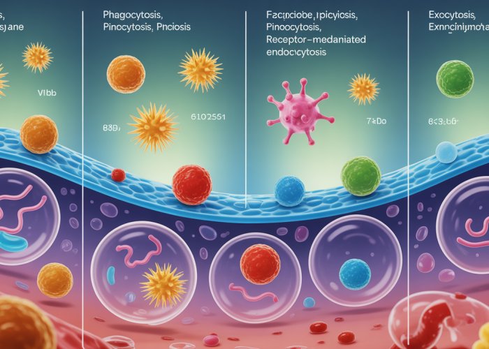
Types of Endocytosis: A Diverse Menu of Uptake Mechanisms
Endocytosis is not a one-size-fits-all process. There are several distinct types, each tailored to internalize specific types of materials or to serve particular cellular functions. These include phagocytosis, pinocytosis, receptor-mediated endocytosis, and caveolae-mediated endocytosis.
Phagocytosis: "Cell Eating" – Engulfing Large Particles
Phagocytosis, literally "cell eating," is the process by which cells engulf large particles, such as bacteria, cellular debris, or even entire dead cells.
It is a crucial defense mechanism employed by specialized cells like macrophages and neutrophils.
The process begins with the recognition of a target particle by receptors on the cell surface. This triggers a cascade of events leading to the formation of pseudopods, temporary extensions of the cell membrane.
These pseudopods reach out and surround the particle, eventually fusing to form a phagosome, a large vesicle containing the engulfed material.
The formation of pseudopods is critically dependent on the cytoskeleton, a network of protein filaments that provides structural support and facilitates cellular movement.
Actin filaments, in particular, play a key role in driving the extension of pseudopods. Once the phagosome is formed, it fuses with a lysosome, an organelle containing digestive enzymes.
The enzymes then break down the engulfed material into smaller molecules that the cell can use.
Pinocytosis: "Cell Drinking" – The Non-Selective Uptake of Extracellular Fluid
In contrast to phagocytosis, which engulfs large particles, pinocytosis, or "cell drinking," is the non-selective uptake of extracellular fluid and its dissolved solutes.
This process involves the invagination of the cell membrane to form small vesicles containing droplets of extracellular fluid.
Pinocytosis occurs continuously in most cell types and serves as a way for cells to sample their environment and take up nutrients.
Unlike receptor-mediated endocytosis, pinocytosis does not require specific receptors to bind to target molecules.
Receptor-Mediated Endocytosis: Highly Specific Uptake of Molecules
Receptor-mediated endocytosis is a highly selective process that allows cells to internalize specific molecules from the extracellular fluid.
This process relies on the presence of receptors on the cell surface that bind to specific target molecules, known as ligands.
Once a ligand binds to its receptor, the receptor-ligand complex migrates to specialized regions of the cell membrane called coated pits.
These pits are coated with a protein called clathrin, which helps to deform the membrane and form a vesicle.
The process of receptor binding, vesicle formation, and uncoating is a carefully orchestrated series of events.
- Receptor Binding: Ligands bind to their specific receptors on the cell surface.
- Vesicle Formation: Clathrin assembles around the receptor-ligand complex, forming a coated pit that invaginates into the cell.
- Uncoating: Once the vesicle is fully formed and pinched off from the cell membrane, the clathrin coat disassembles, leaving a naked vesicle.
The uncoated vesicle then fuses with an endosome, an organelle that sorts and processes internalized materials.
Lysosomes play a critical role in degrading the internalized cargo. Lysosomal enzymes break down the ligands and receptors into their component parts, which can then be recycled by the cell.
Bulk Phase Endocytosis: Non-Selective Entry of Small Molecules
Bulk phase endocytosis is another form of non-selective endocytosis. Similar to pinocytosis, it entraps extracellular fluid.
However, it is characterized by the formation of larger vesicles. This process plays a role in cellular homeostasis.
It helps regulate cell volume and nutrient uptake, although to a lesser extent than the receptor-mediated pathway.
Caveolae: Small Invaginations of the Plasma Membrane
Caveolae are small, flask-shaped invaginations of the plasma membrane that are involved in a variety of cellular processes, including signal transduction, lipid homeostasis, and endocytosis.
These structures are enriched in a protein called caveolin, which is essential for their formation and function.
Caveolae-mediated endocytosis is thought to be involved in the uptake of certain types of molecules, including viruses and toxins.
The exact mechanisms by which caveolae mediate endocytosis are still being investigated.
The Fate of Endocytosed Vesicles: A Journey Through the Cellular Landscape
Once a vesicle is formed through endocytosis, its journey within the cell is far from over. The endocytosed vesicles can take several different paths, each leading to a distinct outcome.
These paths include degradation via lysosomes, recycling back to the cell membrane, or transcytosis.
The Golgi Apparatus plays a crucial role in processing and sorting endocytosed materials. This organelle acts as a central hub for the cell, receiving vesicles from various sources and directing them to their appropriate destinations.
The Golgi modifies, sorts, and packages proteins and lipids, ensuring that they are delivered to the correct locations within the cell.
The pH, or acidity, of the intracellular environment also plays a critical role in endocytosis.
As vesicles move through the endocytic pathway, they encounter progressively more acidic compartments. This change in pH triggers the release of ligands from their receptors, allowing the receptors to be recycled back to the cell membrane.
Implications of Defects in Endocytosis: Diseases and Disorders
Defects in endocytosis can have significant consequences for cellular function and overall health.
Disruptions in endocytic pathways have been implicated in a wide range of diseases and disorders, including cancer, neurodegenerative diseases, and infectious diseases.
For example, impaired receptor-mediated endocytosis can lead to the accumulation of harmful substances in the extracellular space.
Similarly, defects in phagocytosis can compromise the immune system’s ability to clear pathogens and cellular debris.
Understanding the intricacies of endocytosis is crucial for developing new therapies to treat these diseases and disorders.
The internalized vesicles are not simply stored away; they are carefully routed to various intracellular destinations. They may be degraded, recycled, or even transported across the cell to be released on the other side. This intricate system highlights the dynamic nature of the cell membrane and the sophisticated mechanisms that govern cellular import. Now, shifting our focus from the intake process, let’s examine the equally vital mechanism by which cells export materials: exocytosis.
Exocytosis: Cellular Export – Sending Signals and Waste Out
Exocytosis is the fundamental process by which cells release substances into their external environment. Far from being a simple expulsion, it is a meticulously controlled mechanism vital for cellular communication, waste removal, and the delivery of essential molecules.
At its core, exocytosis involves the fusion of intracellular vesicles with the plasma membrane, releasing their contents into the extracellular space. This process is essential for a myriad of cellular functions, from neurotransmitter release in neurons to hormone secretion by endocrine cells.
Types of Exocytosis: A Tale of Two Pathways
Exocytosis is not a monolithic process. It manifests in two primary forms, each serving distinct cellular needs: constitutive exocytosis and regulated exocytosis. Understanding the nuances of each is crucial for appreciating the complexity of cellular export.
Constitutive Exocytosis: The Uninterrupted Flow
Constitutive exocytosis, as the name suggests, is a continuous and unregulated process. It operates in all cells, functioning as a baseline secretion pathway.
This form of exocytosis is primarily responsible for the delivery of newly synthesized membrane proteins and lipids to the plasma membrane.
It also plays a critical role in extracellular matrix (ECM) remodeling and secretion of proteins necessary for maintaining the cell’s structural integrity.
Regulated Exocytosis: On-Demand Release
Regulated exocytosis, in stark contrast to its constitutive counterpart, is a highly controlled process. It occurs only in specialized cells and is triggered by specific signals.
This type of exocytosis is essential for the rapid release of large amounts of specific substances, such as hormones, neurotransmitters, and digestive enzymes.
The triggering signals are diverse, but a common denominator is often an increase in intracellular calcium ion concentration.
The Role of Calcium Ions
Calcium ions (Ca2+) play a pivotal role in triggering regulated exocytosis. Upon receiving a specific stimulus, such as a nerve impulse or a hormonal signal, the intracellular calcium concentration rises sharply.
This surge in calcium ions acts as a signal cascade, initiating a series of events that ultimately lead to the fusion of vesicles with the plasma membrane and the release of their contents.
Vesicle Fusion: The Final Act
The final step in exocytosis is the fusion of the vesicle membrane with the plasma membrane. This fusion event is a complex process that involves the rearrangement of lipids and proteins in both membranes.
The result is the formation of a pore through which the vesicle contents are released into the extracellular space.
Following fusion, the vesicle membrane becomes integrated into the plasma membrane, contributing to its growth and repair.
The Orchestrating Proteins: SNAREs Take Center Stage
Exocytosis is not a spontaneous event. It requires the coordinated action of a diverse array of proteins. Among these, SNARE proteins play a particularly crucial role.
SNAREs (Soluble NSF Attachment Receptor proteins) are a family of proteins that mediate the fusion of vesicles with target membranes. They act like molecular zippers, bringing the vesicle and plasma membranes into close proximity and catalyzing their fusion.
Different SNARE proteins reside on the vesicle (v-SNAREs) and the target membrane (t-SNAREs). Their interaction forms a stable complex that drives membrane fusion, ensuring that exocytosis occurs at the right place and time.
The Golgi Apparatus and Endoplasmic Reticulum: Preparing for Export
The Golgi Apparatus and Endoplasmic Reticulum (ER) play indispensable roles in preparing proteins for exocytosis. These organelles function as the cell’s protein processing and packaging centers.
The ER is the site of protein synthesis and initial folding. From there, proteins destined for secretion are transported to the Golgi Apparatus, where they undergo further modifications, sorting, and packaging into transport vesicles.
These vesicles then bud off from the Golgi and are transported to the plasma membrane for exocytosis.
The Importance of Exocytosis: Beyond Simple Secretion
The significance of exocytosis extends far beyond simply releasing substances from the cell.
It is indispensable for cell communication, enabling cells to transmit signals to one another over short and long distances.
It is also crucial for waste removal, allowing cells to eliminate unwanted materials and maintain cellular homeostasis.
Moreover, exocytosis is vital for membrane protein delivery, ensuring that the plasma membrane has the necessary components to function properly.
In essence, exocytosis is a cornerstone of cellular life, playing a critical role in maintaining cell health, facilitating cell communication, and enabling a wide range of physiological processes.
The release of cellular cargo through exocytosis is just one piece of a much larger and more complex puzzle. Indeed, it’s not enough to simply export substances; the cell must also have a highly organized system for directing traffic within its internal environment. This intricate network, responsible for moving vesicles and other cellular components to their correct destinations, is known as membrane trafficking.
Membrane Trafficking: The Cellular Highway System
Membrane trafficking is the cell’s internal transportation network, a complex and highly regulated system that ensures the correct delivery of proteins, lipids, and other molecules to their appropriate cellular destinations.
Think of it as a sophisticated highway system within the cell, with vesicles acting as the vehicles, transporting cargo along specific routes.
This elaborate system is essential for maintaining cellular organization, enabling communication, and executing a myriad of cellular functions.
The Cytoskeleton: Guiding Vesicle Movement
The cytoskeleton, a dynamic network of protein filaments, serves as the primary infrastructure for membrane trafficking.
Composed of microtubules, actin filaments, and intermediate filaments, the cytoskeleton provides the physical tracks along which vesicles travel.
Motor proteins, such as kinesins and dyneins (associated with microtubules) and myosins (associated with actin filaments), bind to vesicles and use energy from ATP hydrolysis to move them along these tracks.
The selective use of different motor proteins and cytoskeletal tracks allows for precise control over vesicle direction and speed.
This ensures that cargo reaches its intended destination efficiently and accurately.
Furthermore, the cytoskeleton plays a critical role in shaping the cell and facilitating changes in cell shape that are often required for vesicle formation and movement.
Maintaining Cellular Order and Function
The importance of membrane trafficking extends far beyond simple transportation; it is crucial for maintaining cellular organization and overall function.
Disruptions in membrane trafficking can lead to a variety of cellular malfunctions and diseases.
Here’s why:
-
Compartmentalization: Membrane trafficking helps maintain the distinct identities of different cellular compartments, ensuring that each organelle performs its specific functions without interference.
-
Protein and Lipid Sorting: This process allows the cell to sort and deliver newly synthesized proteins and lipids to their correct locations, whether it’s the plasma membrane, Golgi apparatus, or other organelles.
-
Signal Transduction: Membrane trafficking is involved in the transport of receptors and signaling molecules, playing a key role in cellular communication and responses to external stimuli.
-
Nutrient Uptake and Waste Removal: Both endocytosis and exocytosis, integral parts of membrane trafficking, are essential for nutrient uptake and the removal of waste products, maintaining cellular homeostasis.
In essence, membrane trafficking is not just about moving things around; it’s about ensuring that the cell functions as a well-oiled machine, with each component working in harmony to maintain its overall health and viability.
Without a properly functioning membrane trafficking system, the cell would quickly descend into chaos, unable to maintain its structure, communicate effectively, or respond appropriately to its environment.
The release of cellular cargo through exocytosis is just one piece of a much larger and more complex puzzle. Indeed, it’s not enough to simply export substances; the cell must also have a highly organized system for directing traffic within its internal environment. This intricate network, responsible for moving vesicles and other cellular components to their correct destinations, is known as membrane trafficking. Now, consider that everything brought into the cell via endocytosis must also be carefully managed, sorted, and perhaps even expelled. It’s here, at the intersection of import and export, that we begin to appreciate the elegant dance between endocytosis and exocytosis.
The Interplay of Endocytosis and Exocytosis: A Harmonious Balance
Endocytosis and exocytosis, at first glance, might appear as opposing forces – one bringing materials into the cell, the other expelling them.
However, a closer examination reveals a far more nuanced relationship; these processes are not merely opposing, but are instead intricately coordinated to maintain cellular equilibrium and facilitate critical functions.
Indeed, their cooperative action is essential for maintaining cell membrane homeostasis and enabling effective cellular communication.
Maintaining Cell Membrane Homeostasis: A Balancing Act
The cell membrane is a dynamic structure, constantly undergoing remodeling and turnover.
Endocytosis removes portions of the plasma membrane, internalizing them as vesicles.
Conversely, exocytosis adds membrane back to the cell surface through vesicle fusion.
This continuous cycle of membrane retrieval and insertion is crucial for maintaining a stable cell size and surface area.
Without this balance, cells would either shrink due to excessive endocytosis or expand uncontrollably due to unchecked exocytosis.
This dynamic equilibrium ensures that the cell membrane retains its structural integrity and functional capacity.
Moreover, the lipid and protein composition of the plasma membrane must remain relatively constant for proper cellular function.
The coordinated action of endocytosis and exocytosis helps to maintain this balance by regulating the insertion and removal of specific membrane components.
For instance, receptors internalized via endocytosis can be either degraded or recycled back to the cell surface via exocytosis, allowing cells to adjust their sensitivity to external signals.
This intricate interplay ensures that the cell membrane remains optimally configured to meet the cell’s ever-changing needs.
Signal Transduction and Cellular Communication: A Two-Way Street
Endocytosis and exocytosis play critical roles in cell signaling, enabling cells to communicate with their environment and with each other.
Receptor-mediated endocytosis, for example, is a key mechanism for initiating and regulating signaling cascades.
Upon binding to their ligands at the cell surface, receptors are internalized via endocytosis, triggering downstream signaling events.
This process not only allows cells to respond to external stimuli but also provides a means of modulating the duration and intensity of signaling.
Furthermore, endocytosis can terminate signaling by removing receptors from the cell surface, preventing overstimulation.
Exocytosis, on the other hand, is essential for releasing signaling molecules, such as neurotransmitters and hormones, into the extracellular space.
This allows cells to communicate with neighboring cells or distant tissues, coordinating physiological responses throughout the organism.
The regulated secretion of these molecules through exocytosis is tightly controlled, ensuring that signals are delivered at the right time and to the right location.
Moreover, exocytosis can also deliver newly synthesized receptors to the cell surface, replenishing the receptor pool and allowing cells to maintain their responsiveness to signals.
In essence, endocytosis and exocytosis function as a two-way communication system, enabling cells to both receive and transmit information, orchestrating complex cellular behaviors and physiological processes.
The disruption of this delicate balance can have profound consequences, leading to a variety of diseases and disorders.
Endocytosis & Exocytosis FAQs
Here are some frequently asked questions about endocytosis and exocytosis, the processes cells use to transport materials in and out.
What’s the main difference between endocytosis and exocytosis?
Endocytosis brings substances into the cell by engulfing them in a vesicle formed from the cell membrane. Exocytosis, conversely, releases substances out of the cell by fusing a vesicle containing the substance with the cell membrane. Essentially, endocytosis imports, and exocytosis exports.
What are some specific examples of substances transported by endocytosis?
Endocytosis is used to transport a wide range of materials, including nutrients, hormones, pathogens, and even entire cells in some cases. For example, immune cells use endocytosis to engulf and destroy bacteria. The cell membrane creates a pocket around the particle, then seals off.
What are the different types of endocytosis?
There are several types of endocytosis, including phagocytosis (cell eating), pinocytosis (cell drinking), and receptor-mediated endocytosis, which is more specific. Each type utilizes a slightly different mechanism to internalize materials.
Why are endocytosis and exocytosis important for cell survival?
Endocytosis and exocytosis are vital for cell survival because they enable cells to maintain their internal environment and communicate with their surroundings. These processes allow cells to acquire essential nutrients, eliminate waste products, and secrete hormones or enzymes, all crucial for proper functioning.
So, that’s a wrap on endocytosis and exocytosis! Hopefully, you now have a better grasp of these amazing cellular processes. Now go forth and share your newfound knowledge!