Understanding the intricate rules of DNA is fundamental for advancements in fields like genetic engineering. The Watson-Crick model serves as the cornerstone, illustrating how nucleotides pair to form the iconic double helix structure. Further, the National Institutes of Health (NIH) contributes significantly to unraveling these complexities through extensive research initiatives. Ultimately, these discoveries directly impact the work of scientists like Jennifer Doudna, enabling precise gene editing and furthering our grasp of the underlying rules of DNA and its implications for health and disease.
Deoxyribonucleic acid, or DNA, is the fundamental molecule that carries the genetic instructions for all known living organisms and many viruses. It’s the very essence of life, dictating everything from the color of our eyes to our susceptibility to certain diseases.
Understanding DNA is paramount to understanding life itself.
The Importance of DNA in Biology
DNA’s importance stems from its role as the blueprint for life. It contains the instructions needed for an organism to develop, survive, and reproduce. Without DNA, there would be no inheritance, no evolution, and ultimately, no life as we know it.
DNA is not merely a static archive of information.
It is a dynamic molecule that interacts with its environment, responds to signals, and orchestrates the complex processes within cells.
Why the "Rules of DNA" Matter
Comprehending the "rules of DNA" is crucial for deciphering the intricate mechanisms that govern life processes. These rules dictate how DNA is structured, how it replicates, how it’s transcribed into RNA, and ultimately, how it translates into proteins.
These proteins then carry out the vast majority of cellular functions.
By understanding these rules, we can gain insights into:
- The causes of genetic diseases.
- The development of new therapies.
- The evolution of life on Earth.
Purpose of This Article
This article aims to provide a comprehensive explanation of the fundamental principles governing DNA’s structure, function, and behavior.
We will explore the key concepts that underpin our understanding of this remarkable molecule, from its double helix structure to the intricate processes of replication, transcription, and translation.
By the end of this article, you will have a solid foundation in the "rules of DNA," equipping you with the knowledge to appreciate the complexity and beauty of the genetic code.
Deoxyribonucleic acid, or DNA, is the fundamental molecule that carries the genetic instructions for all known living organisms and many viruses. It’s the very essence of life, dictating everything from the color of our eyes to our susceptibility to certain diseases. Understanding DNA is paramount to understanding life itself.
DNA’s importance stems from its role as the blueprint for life. It contains the instructions needed for an organism to develop, survive, and reproduce. Without DNA, there would be no inheritance, no evolution, and ultimately, no life as we know it.
DNA is not merely a static archive of information. It is a dynamic molecule that interacts with its environment, responds to signals, and orchestrates the complex processes within cells.
Comprehending the "rules of DNA" is crucial for deciphering the intricate mechanisms that govern life processes. These rules dictate how DNA is structured, how it replicates, how it’s transcribed into RNA, and ultimately, how it translates into proteins. These proteins then carry out the vast majority of cellular functions.
By understanding these rules, we can gain insights into:
- The causes of genetic diseases.
- The development of new therapies.
- The evolution of life on Earth.
This article aims to provide a comprehensive explanation of the fundamental principles governing DNA’s structure, function, and behavior. We will explore the key concepts that underpin our understanding of this remarkable molecule, from its double helix structure to the intricate processes that govern its activity.
But before we can delve into the intricate processes that DNA governs, we must first understand its fundamental structure. After all, the architecture of a molecule often dictates its function. Let’s begin to uncover the foundational building blocks of this essential molecule, the very blueprint of life itself.
The Foundation: Decoding DNA Structure
DNA, the molecule that holds the key to our genetic code, is renowned for its elegant and distinctive double helix structure. But what exactly does this structure entail, and what are the components that make up this vital molecule? Let’s break down the architecture of DNA, examining its building blocks and how they assemble to form the iconic double helix.
Unraveling the Double Helix
Imagine a twisted ladder, that’s the essence of the DNA’s double helix. Two strands of DNA wind around each other, forming a spiral staircase-like shape.
These strands are not identical but complementary, fitting together in a precise and predictable manner. This double helix structure is not just aesthetically pleasing; it’s fundamental to DNA’s ability to store and transmit genetic information.
The beauty of the double helix lies in its stability and its ability to be easily replicated, ensuring that genetic information is passed on accurately from one generation to the next.
The Components of a Nucleotide
The building blocks of DNA are called nucleotides. Each nucleotide consists of three essential components:
- A deoxyribose sugar: This is a five-carbon sugar molecule that forms the backbone of the DNA strand.
- A phosphate group: This group is attached to the deoxyribose sugar and provides the linkage between nucleotides, forming the sugar-phosphate backbone of the DNA strand.
- A nitrogenous base: This is the information-carrying component of the nucleotide, and it’s what distinguishes one nucleotide from another.
These three components link together to form a single nucleotide, and many nucleotides join to create a strand of DNA. The sugar-phosphate backbone provides structural support, while the nitrogenous bases encode the genetic information.
The Four Nitrogenous Bases
There are four types of nitrogenous bases found in DNA:
- Adenine (A)
- Guanine (G)
- Cytosine (C)
- Thymine (T)
These bases are categorized into two groups: purines (Adenine and Guanine), which have a double-ring structure, and pyrimidines (Cytosine and Thymine), which have a single-ring structure. The sequence of these bases along the DNA strand determines the genetic code.
The specific order of these bases dictates the genetic instructions encoded within the DNA molecule. It’s this sequence that ultimately determines the characteristics of an organism. Therefore, understanding these bases is fundamental to understanding how DNA functions.
But before we can delve into the intricate processes that DNA governs, it’s crucial to grasp one of its most fundamental principles – the concept of base pairing. This seemingly simple rule holds the key to understanding DNA’s stability, replication, and overall function.
The Golden Rule: Mastering Base Pairing
At the heart of DNA’s functionality lies the elegant and precise rule of base pairing. This principle dictates how the nitrogenous bases within the DNA molecule interact, forming the "rungs" of the DNA ladder and ensuring the molecule’s structural integrity. Understanding this "golden rule" is paramount to unlocking the secrets of DNA.
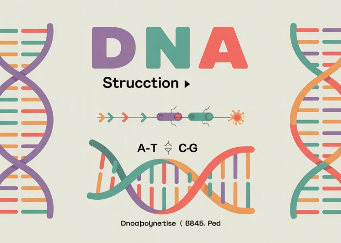
The A-T and G-C Partnerships
The base pairing rule is straightforward yet profoundly significant: Adenine (A) always pairs with Thymine (T), and Guanine (G) always pairs with Cytosine (C).
This specificity isn’t arbitrary; it’s dictated by the molecular structure of the bases and their ability to form stable hydrogen bonds.
Adenine and Thymine form two hydrogen bonds, while Guanine and Cytosine form three. This difference in hydrogen bonding contributes to the overall stability of the DNA double helix.
This strict adherence to pairing ensures that the two strands of DNA are complementary to each other.
Meaning, knowing the sequence of one strand automatically reveals the sequence of the other.
Hydrogen Bonds: The Glue That Holds It All Together
The stability of the DNA double helix isn’t solely dependent on the base pairing itself; the hydrogen bonds that form between the paired bases play a crucial role.
These bonds, though individually weak, collectively provide the strength needed to maintain the double helix structure.
Hydrogen bonds act like tiny molecular "glue," holding the two DNA strands together.
Without these bonds, the DNA molecule would unravel and lose its structural integrity. The two hydrogen bonds between A and T and three hydrogen bonds between G and C is the precise amount needed to create stability.
The Profound Significance of Accurate Base Pairing
The seemingly simple rule of base pairing has far-reaching implications for DNA replication, transcription, and ultimately, the accurate transmission of genetic information.
Ensuring Faithful DNA Replication
During DNA replication, the double helix unwinds, and each strand serves as a template for the synthesis of a new complementary strand.
The base pairing rules ensure that the newly synthesized strands are accurate copies of the original DNA molecule.
If A didn’t always pair with T, and G with C, the replicated DNA would contain errors, leading to mutations and potentially harmful consequences.
Guiding Transcription and RNA Synthesis
Similarly, base pairing is crucial for transcription, the process of creating RNA from a DNA template.
RNA polymerase, the enzyme responsible for transcription, uses the base pairing rules to synthesize an RNA molecule that is complementary to the DNA template strand.
However, instead of Thymine (T), RNA uses Uracil (U), which pairs with Adenine (A). This difference in base composition is key to distinguishing RNA from DNA and allows for the proper functioning of each molecule.
Maintaining Genetic Integrity
In essence, the accuracy of base pairing ensures the integrity of the genetic code.
By adhering to the A-T and G-C rule, DNA can be faithfully replicated and transcribed, preserving the genetic information necessary for life.
Any deviation from this rule can lead to mutations, which can have a variety of effects, ranging from no noticeable change to severe genetic disorders.
Therefore, mastering the base pairing rule is not just about memorizing a simple principle; it’s about understanding the foundation upon which all of molecular biology is built.
The hydrogen bonds between base pairs are indeed the glue that holds the double helix together, but they are not the whole story. The architecture of DNA depends on the faithful duplication of the original strand; this must occur with each cell division so that the new cells also possess the exact same genome.
Copying the Code: Understanding DNA Replication
At its core, DNA replication is the fundamental process by which a cell duplicates its DNA. This ensures that each daughter cell receives an identical copy of the genetic material. Imagine trying to copy a massive, intricate blueprint by hand. The potential for errors would be immense. Yet, cells manage this feat with astonishing accuracy.
The Semiconservative Nature of Replication
DNA replication follows a semiconservative model. This means that each new DNA molecule consists of one original (template) strand and one newly synthesized strand. This elegantly preserves the integrity of the genetic information. Think of it as making a copy of a document while keeping the original safe.
The Replication Fork: Where the Action Happens
The process begins at specific locations on the DNA molecule called origins of replication. Here, the double helix unwinds, creating a "replication fork"—a Y-shaped structure where the DNA strands separate.
This unwinding is facilitated by enzymes called helicases. These act like molecular zippers, breaking the hydrogen bonds between base pairs.
DNA Polymerase: The Master Builder
The star of the show is DNA polymerase, an enzyme that catalyzes the synthesis of new DNA strands. DNA polymerase can only add nucleotides to the 3′ (three prime) end of an existing strand.
This directionality has significant implications for how replication proceeds on the two template strands.
Leading and Lagging Strands: A Tale of Two Syntheses
Because DNA polymerase can only add nucleotides in the 5′ to 3′ direction, replication occurs differently on the two strands. The leading strand is synthesized continuously in the 5′ to 3′ direction as the replication fork opens.
The lagging strand, however, is synthesized discontinuously. It creates short fragments called Okazaki fragments, also in the 5′ to 3′ direction.
These fragments are later joined together by another enzyme called DNA ligase.
Primers: Getting the Process Started
DNA polymerase cannot initiate DNA synthesis on its own. It requires a short RNA sequence called a primer to get started.
This primer is synthesized by an enzyme called primase. Once the primer is in place, DNA polymerase can then add nucleotides to the 3′ end.
Ensuring Accuracy: Proofreading and Repair Mechanisms
DNA replication is remarkably accurate, but errors can still occur.
DNA polymerase has a proofreading function. It can recognize and remove incorrectly added nucleotides during replication.
Furthermore, cells have various DNA repair mechanisms to correct any errors that escape proofreading.
These mechanisms involve enzymes that scan the DNA for damage, remove the damaged section, and then use the undamaged strand as a template to synthesize the correct sequence.
Maintaining Genetic Integrity: The Importance of Accurate Replication
The accuracy of DNA replication is paramount. Even a small error rate can have devastating consequences. Mutations can lead to a variety of problems, including genetic disorders and cancer.
Therefore, the intricate mechanisms that ensure accurate DNA replication are essential for maintaining the integrity of the genome and the health of the organism.
The meticulous process of DNA replication ensures the faithful inheritance of genetic information. However, the information encoded within DNA must also be accessed and utilized to direct cellular activities. This is where the elegant process of transcription comes into play, serving as the crucial link between the genetic blueprint and the cellular machinery.
From DNA to RNA: The Process of Transcription
Transcription is the cellular mechanism by which the information encoded in DNA is copied into a complementary RNA molecule. This RNA molecule then serves as a template for protein synthesis or plays other functional roles within the cell. Think of it as creating a working copy of a master document, a copy that can be directly used without risking damage to the original.
The Central Role of RNA Polymerase
The enzyme responsible for carrying out transcription is RNA polymerase. Unlike DNA polymerase, RNA polymerase does not require a primer to initiate synthesis. Instead, it binds to specific DNA sequences called promoters, which signal the starting point for transcription.
RNA polymerase then unwinds the DNA double helix and begins synthesizing an RNA molecule complementary to one of the DNA strands, known as the template strand.
The RNA molecule is synthesized in the 5′ to 3′ direction, adding nucleotides to the 3′ end of the growing chain. The sequence of the RNA molecule is determined by the base pairing rules: adenine (A) pairs with uracil (U) in RNA, guanine (G) pairs with cytosine (C), and vice versa.
Messenger, Transfer, and Ribosomal RNA
Transcription produces different types of RNA molecules, each with its own specific function. The three major types are:
-
Messenger RNA (mRNA): mRNA carries the genetic information from DNA to the ribosomes, where it is translated into protein. Each mRNA molecule contains a sequence of codons, each of which specifies a particular amino acid in the protein sequence.
-
Transfer RNA (tRNA): tRNA molecules act as adaptors, bringing the correct amino acid to the ribosome during translation. Each tRNA molecule has an anticodon that recognizes a specific codon on the mRNA molecule.
-
Ribosomal RNA (rRNA): rRNA is a major component of ribosomes, the cellular structures where protein synthesis takes place. rRNA molecules play both structural and catalytic roles in the ribosome.
Uracil: The Key Difference
One of the key differences between DNA and RNA is the presence of uracil (U) in RNA instead of thymine (T) in DNA. Uracil is a pyrimidine base that is similar in structure to thymine, but it lacks a methyl group.
In RNA, uracil pairs with adenine (A), just as thymine does in DNA. The presence of uracil in RNA allows it to form different secondary structures compared to DNA, which is important for its various functional roles.
Uracil’s slightly different structure also makes RNA less stable than DNA, which is appropriate for its role as a temporary carrier of genetic information.
RNA to Protein: Decoding Translation
With the creation of mRNA through transcription, the cell now holds a working copy of the genetic instructions. This mRNA molecule, however, is not the final product. It’s merely a messenger, carrying the code that will ultimately be translated into the functional units of the cell: proteins. The process of translation is where this remarkable conversion occurs, bringing the genetic code to life.
The Ribosome: The Protein Synthesis Workhorse
The key player in translation is the ribosome, a complex molecular machine found in all living cells. Ribosomes are composed of two subunits, a large subunit and a small subunit, which come together to facilitate protein synthesis.
Think of the ribosome as a construction site where proteins are built.
It provides the necessary framework and enzymatic activity to link amino acids together in the correct order, as dictated by the mRNA sequence.
tRNA: The Adapter Molecule
Transfer RNA (tRNA) molecules act as adapter molecules, bridging the gap between the nucleotide sequence of the mRNA and the amino acid sequence of the protein.
Each tRNA molecule has a specific anticodon sequence that is complementary to a particular codon on the mRNA. It also carries the amino acid corresponding to that codon.
This ensures that the correct amino acid is added to the growing polypeptide chain.
Codons: The Language of Life
The genetic code is read in three-nucleotide units called codons. Each codon specifies a particular amino acid, or a start/stop signal.
For example, the codon AUG signals the start of translation and also codes for the amino acid methionine.
There are 64 possible codons, but only 20 amino acids commonly found in proteins. This redundancy means that some amino acids are specified by more than one codon.
This redundancy is thought to provide some protection against the effects of mutations.
The Translation Process: A Step-by-Step Guide
Translation can be divided into three main stages: initiation, elongation, and termination.
Initiation
Initiation begins with the small ribosomal subunit binding to the mRNA and a special initiator tRNA carrying methionine. The complex moves along the mRNA until it finds the start codon (AUG). Then, the large ribosomal subunit joins, forming the complete ribosome complex.
Elongation
During elongation, the ribosome moves along the mRNA, one codon at a time. For each codon, a tRNA molecule with the corresponding anticodon binds to the mRNA, delivering its amino acid.
The ribosome then catalyzes the formation of a peptide bond between the incoming amino acid and the growing polypeptide chain. The "empty" tRNA molecule then exits the ribosome.
This process repeats as the ribosome continues to move along the mRNA.
Termination
Termination occurs when the ribosome encounters a stop codon (UAA, UAG, or UGA) on the mRNA. These codons do not code for any amino acid.
Instead, they signal the end of translation. Release factors bind to the ribosome, causing the polypeptide chain to be released and the ribosome to dissociate into its subunits.
From Codon to Protein: The Precision of the Process
The sequence of codons in the mRNA directly determines the sequence of amino acids in the protein.
This sequence is critical because the amino acid sequence dictates the protein’s three-dimensional structure and, therefore, its function.
A single change in the codon sequence can result in a different amino acid being incorporated into the protein, which can alter its structure and function.
This highlights the exquisite precision of translation and the importance of maintaining the integrity of the genetic code.
RNA’s journey from a transcribed message to a functional protein highlights the elegance of cellular processes. But what exactly dictates which amino acid is added to the growing polypeptide chain based on the mRNA sequence? This is where the genetic code comes into play, acting as the Rosetta Stone for translating nucleotide language into the language of proteins.
The Genetic Code: Cracking the Cellular Language
The genetic code is the complete set of rules used by living cells to translate information encoded in genetic material (DNA or RNA sequences) into proteins. It’s a dictionary, matching three-letter "words" (codons) in the mRNA to specific amino acids.
Deciphering the Codon
Each codon consists of a sequence of three nucleotides, representing a specific amino acid or a start/stop signal during translation. With four possible nucleotides (A, U, G, C) at each of the three positions, there are 64 possible codons (4 x 4 x 4 = 64).
For example, the codon AUG not only codes for the amino acid methionine but also acts as the start codon, initiating protein synthesis. Conversely, codons like UAA, UAG, and UGA are stop codons, signaling the termination of translation and the release of the newly synthesized polypeptide chain.
Redundancy: Multiple Codons for a Single Amino Acid
The genetic code is described as degenerate or redundant, meaning that multiple codons can code for the same amino acid. This redundancy is not uniform; some amino acids are specified by only one codon, while others are specified by as many as six.
The wobble hypothesis suggests that the third nucleotide in a codon is less critical in determining the amino acid, allowing for some flexibility in tRNA binding. This degeneracy can provide a buffer against mutations, as a change in the third nucleotide of a codon may still result in the same amino acid being incorporated into the protein.
Universality: A Shared Language of Life
One of the most remarkable features of the genetic code is its near universality. With few exceptions, the same codons specify the same amino acids in virtually all organisms, from bacteria to humans.
This universality provides compelling evidence for the common ancestry of all life on Earth. It also allows for the possibility of transferring genetic information between different organisms, a cornerstone of genetic engineering and biotechnology. While minor variations exist in the mitochondrial genetic code and in certain organisms, the core principles remain remarkably conserved.
Understanding the genetic code is critical for deciphering the functional consequences of gene sequences and mutations. It provides the foundation for understanding how cells synthesize proteins and how genetic information is inherited and expressed.
RNA’s journey from a transcribed message to a functional protein highlights the elegance of cellular processes. But what exactly dictates which amino acid is added to the growing polypeptide chain based on the mRNA sequence? This is where the genetic code comes into play, acting as the Rosetta Stone for translating nucleotide language into the language of proteins.
Central Dogma of Molecular Biology: The Flow of Genetic Information
The central dogma of molecular biology is a cornerstone concept, encapsulating the fundamental flow of genetic information within biological systems. It elegantly describes how information, encoded in DNA, is ultimately expressed as functional proteins, the workhorses of the cell.
At its core, the central dogma outlines a sequential process: from DNA to RNA to protein. This unidirectional flow, while not without exceptions, provides a powerful framework for understanding gene expression and cellular function.
DNA Replication: Preserving the Genetic Blueprint
The process begins with DNA replication, a critical step ensuring the faithful duplication of the genetic material. This is essential for cell division and the transmission of genetic information to subsequent generations.
During replication, the double helix unwinds, and each strand serves as a template for the synthesis of a new, complementary strand. The result is two identical DNA molecules, each containing one original and one newly synthesized strand.
This semi-conservative replication is orchestrated by enzymes like DNA polymerase, which precisely adds nucleotides to the growing strand, adhering to the base pairing rules (A with T, G with C).
RNA Transcription: Copying the Message
Next, transcription involves the creation of an RNA molecule from a DNA template. This process is mediated by RNA polymerase, which binds to specific DNA sequences and synthesizes a complementary RNA strand.
Unlike DNA replication, transcription doesn’t copy the entire DNA molecule. Instead, it selectively transcribes specific genes, creating RNA molecules that carry the instructions for protein synthesis.
There are different types of RNA, each with a specific role. Messenger RNA (mRNA) carries the genetic code from DNA to ribosomes, the protein synthesis machinery. Transfer RNA (tRNA) brings amino acids to the ribosome, matching them to the codons on the mRNA. Ribosomal RNA (rRNA) is a structural component of ribosomes.
Protein Translation: Decoding the Instructions
Finally, translation uses the information encoded in mRNA to synthesize proteins. This process occurs at the ribosomes, where mRNA is read in three-nucleotide units called codons.
Each codon corresponds to a specific amino acid, and tRNA molecules, carrying the appropriate amino acids, bind to the mRNA codon, delivering their amino acid cargo.
As the ribosome moves along the mRNA, amino acids are linked together, forming a growing polypeptide chain. This chain folds into a specific three-dimensional structure, resulting in a functional protein.
Exceptions to the Rule: Expanding the Dogma
While the central dogma provides a valuable framework, it is important to acknowledge that there are exceptions.
For example, retroviruses, like HIV, can reverse transcribe RNA into DNA, defying the typical flow of information. Additionally, some RNA molecules can directly regulate gene expression without being translated into protein.
These exceptions highlight the complexity and adaptability of biological systems, reminding us that the central dogma is not an immutable law but a guiding principle that helps us understand the flow of genetic information.
When Errors Occur: Exploring Mutations
The fidelity of DNA replication and transcription is remarkably high, yet it’s not perfect. Occasionally, alterations in the DNA sequence arise, known as mutations. These mutations, though seemingly small changes at the molecular level, can have profound consequences, shaping the course of evolution and influencing individual health.
Defining Mutations: Alterations in the Genetic Code
At its core, a mutation is defined as any change in the nucleotide sequence of DNA. This alteration can range from a single base pair substitution to larger-scale changes involving entire sections of a chromosome.
Mutations can occur spontaneously during DNA replication, or they can be induced by external factors such as radiation, chemicals, or viruses.
The permanency of the changes caused by mutations is what distinguishes them from temporary changes.
These changes can then be passed to future generations, if they occur in germline cells (sperm or egg cells).
Types of Mutations: A Diverse Landscape of Genetic Change
Mutations are not a monolithic entity; they manifest in various forms, each with its unique mechanism and potential impact:
-
Point Mutations: These are the most common type of mutation, involving a change in a single nucleotide base. There are several types of point mutations:
-
Substitutions: One base is replaced by another. For example, an adenine (A) might be replaced by a guanine (G). These substitutions can be further classified as:
- Transitions: A purine (A or G) is replaced by another purine, or a pyrimidine (C or T) is replaced by another pyrimidine.
- Transversions: A purine is replaced by a pyrimidine, or vice versa.
-
Insertions: One or more nucleotide bases are added to the DNA sequence.
-
Deletions: One or more nucleotide bases are removed from the DNA sequence.
-
-
Frameshift Mutations: Insertions or deletions can be particularly disruptive if they involve a number of bases that is not a multiple of three.
This is because the genetic code is read in triplets (codons), each codon specifying an amino acid.Adding or deleting bases shifts the reading frame, altering the sequence of amino acids and potentially leading to a nonfunctional protein.
-
Chromosomal Mutations: These are large-scale mutations affecting entire chromosomes or significant portions thereof. They include:
- Deletions: Loss of a portion of a chromosome.
- Duplications: Replication of a portion of a chromosome, resulting in multiple copies of the same region.
- Inversions: A segment of a chromosome is reversed end-to-end.
- Translocations: A segment of a chromosome breaks off and attaches to another chromosome.
Consequences of Mutations: A Spectrum of Effects
The impact of a mutation can vary widely, depending on the location and nature of the change. Some mutations have no discernible effect, while others can be detrimental or even beneficial.
-
Harmful Mutations: Many mutations are harmful, disrupting normal cellular function and leading to disease.
- Mutations in genes that regulate cell growth and division can lead to uncontrolled cell proliferation and cancer.
- Mutations in genes encoding essential proteins can cause genetic disorders such as cystic fibrosis, sickle cell anemia, and Huntington’s disease.
-
Beneficial Mutations: Although less common, some mutations can be beneficial, providing an organism with a selective advantage.
- For example, a mutation that confers resistance to a particular disease could increase an organism’s survival rate in an environment where that disease is prevalent.
- Beneficial mutations are the driving force behind evolution, allowing organisms to adapt to changing environments over time.
-
Neutral Mutations: These mutations have no significant effect on the organism’s phenotype.
- They often occur in non-coding regions of DNA or result in a change in the amino acid sequence of a protein that does not alter its function.
- Neutral mutations can accumulate over time and contribute to genetic diversity within a population.
Mutations are a double-edged sword. They are a source of genetic variation, the raw material for evolution, but they can also lead to disease and disability. Understanding the nature and consequences of mutations is crucial for advancing our knowledge of biology and medicine, from developing new therapies for genetic disorders to understanding the evolution of life on Earth.
Genes and Chromosomes: Organizing the Genetic Material
Having explored the world of mutations and their impact on the genetic code, it’s important to zoom out and examine the larger organizational context of DNA. How is all this genetic information packaged and arranged within the cell? The answer lies in genes and chromosomes, the fundamental units of heredity and the structures that house them.
Defining the Gene: The Functional Unit of Heredity
At its most basic, a gene is a specific sequence of DNA that contains the instructions for building a protein or RNA molecule. Think of it as a blueprint for a particular cellular product.
These products, proteins and functional RNA molecules, carry out a vast array of functions within the cell, from catalyzing biochemical reactions to providing structural support.
Some genes encode proteins that serve as enzymes, hormones, or structural components, while others code for functional RNA molecules like transfer RNA (tRNA) or ribosomal RNA (rRNA), which play crucial roles in protein synthesis.
Therefore, each gene contributes to an organism’s traits, from eye color to susceptibility to certain diseases.
Chromosomes: Organizing Genes into Manageable Units
If genes are the individual blueprints, then chromosomes are the filing cabinets that organize and store them.
Chromosomes are thread-like structures composed of DNA tightly wound around proteins called histones. This compact packaging is essential for fitting the enormous amount of DNA within the limited space of the cell nucleus.
Chromosome Structure and Function
The structure of a chromosome is not uniform throughout its length. There are regions of tightly packed DNA, called heterochromatin, that are generally inactive in terms of gene expression.
Conversely, euchromatin is more loosely packed and contains genes that are actively being transcribed.
Each chromosome has a centromere, a constricted region that plays a critical role in cell division.
Telomeres, protective caps at the ends of chromosomes, prevent DNA degradation and maintain chromosomal stability.
Genes Organized on Chromosomes
Genes are arranged linearly along each chromosome, with each gene occupying a specific location, or locus. The order and arrangement of genes on a chromosome are highly conserved, meaning that they tend to remain relatively stable over evolutionary time.
This organization ensures that genes are inherited in a predictable manner from one generation to the next.
The Human Genome: A Vast and Complex Library
The human genome is the complete set of DNA found within the 23 pairs of chromosomes in the nucleus of a human cell.
It represents the totality of our genetic inheritance, containing an estimated 20,000 to 25,000 protein-coding genes. However, it is important to note that genes make up only a small fraction of the total human genome.
Unraveling the Mysteries of Non-Coding DNA
Much of the human genome consists of non-coding DNA, sequences that do not directly code for proteins.
While initially dismissed as "junk DNA," it is now recognized that non-coding DNA plays critical roles in gene regulation, chromosome structure, and other essential cellular processes.
These non-coding regions can include regulatory elements that control when and where genes are expressed, as well as structural elements that help maintain chromosome integrity.
The Dynamic Nature of the Genome
The human genome is not a static entity. It is constantly being reshaped by mutations, recombination, and other dynamic processes.
Understanding the organization and complexity of the human genome is crucial for unraveling the mysteries of human biology and disease.
By studying the interplay between genes, chromosomes, and the non-coding regions of the genome, scientists are gaining new insights into the fundamental processes that govern life. This knowledge will pave the way for the development of new diagnostic tools, therapies, and strategies for improving human health.
The Discoverers: Recognizing Key Figures in DNA Research
The unraveling of DNA’s structure stands as a monumental achievement in scientific history. However, like many great discoveries, the path to understanding was paved by the contributions—and sometimes, the overlooked contributions—of several brilliant minds. While James Watson and Francis Crick are often credited with the discovery, it’s crucial to acknowledge the vital role of Rosalind Franklin, whose experimental work provided critical insights.
The Central Contributions
Rosalind Franklin: The Unsung Heroine
Rosalind Franklin’s work at King’s College London was pivotal in revealing the structure of DNA. Her X-ray diffraction images, most notably "Photo 51," provided crucial data about the molecule’s helical nature. This image, in particular, offered key measurements and insights that were instrumental in deducing the double helix structure.
It is essential to acknowledge that Franklin’s contributions were not always appropriately recognized during her lifetime. Her meticulous experimental work, rigorous analysis, and sharp intellect laid a crucial foundation for understanding DNA.
James Watson and Francis Crick: The Model Builders
James Watson and Francis Crick, working at the University of Cambridge, are widely recognized for building the first accurate model of the DNA double helix. Their approach involved synthesizing existing research, including Franklin’s X-ray diffraction data and Erwin Chargaff’s base pairing rules (A with T, and G with C).
They ingeniously combined these insights to propose a structure that elegantly explained DNA’s ability to store and transmit genetic information. While Watson and Crick are celebrated for their model, the reliance on Franklin’s data underscores the collaborative nature of scientific progress—and the importance of acknowledging all contributors.
The Impact on Our Understanding of Genetics
The elucidation of DNA’s structure revolutionized our understanding of genetics. It provided a physical basis for how genetic information is stored, replicated, and passed on to future generations. This breakthrough paved the way for countless advancements in medicine, biotechnology, and other fields.
A Foundation for Modern Biology
Understanding DNA’s structure has enabled scientists to develop techniques for gene cloning, DNA sequencing, and genetic engineering. These tools have had a profound impact on our ability to diagnose and treat diseases, develop new therapies, and improve crop yields.
Ethical Considerations and Legacy
The story of DNA’s discovery also raises important ethical considerations about scientific collaboration and recognition. The fact that Rosalind Franklin’s contributions were not fully acknowledged until after her death serves as a reminder of the challenges faced by women in science during that era.
Acknowledging the contributions of all scientists involved, including those whose work may have been overlooked, is essential for promoting equity and integrity in the scientific community. It ensures that future generations of scientists learn from the past and strive to create a more inclusive and collaborative environment.
Decoding DNA: Frequently Asked Questions
This FAQ section addresses common questions about decoding DNA and the important concepts discussed in the article. Hopefully, it will help clarify any confusion and further your understanding.
What does it mean to "decode" DNA?
Decoding DNA essentially means understanding the sequence of nucleotides (A, T, C, and G) and how that sequence translates into the instructions for building proteins. It involves deciphering the genetic code based on the established rules of DNA, which determines what proteins are made and when.
What are the key "rules of DNA" needed for decoding?
The core rules of DNA include understanding the complementary base pairing (A with T, and C with G), the triplet codon system (three nucleotides code for one amino acid), and the start and stop codons that initiate and terminate protein synthesis. These rules of DNA allow us to translate the genetic code.
How do scientists "read" the DNA sequence?
Scientists use techniques like DNA sequencing to determine the exact order of nucleotides in a DNA molecule. Once the sequence is known, computational tools and databases are used to identify genes, predict protein sequences, and understand the function of those proteins.
Why is understanding DNA decoding important?
Understanding DNA decoding is crucial for many areas, including medicine, agriculture, and biotechnology. It helps us understand genetic diseases, develop new therapies, improve crop yields, and engineer organisms for specific purposes. Applying the rules of DNA unlocks countless possibilities.
So, there you have it! Hopefully, this deep dive into the **rules of DNA** has clarified things. Now go forth and impress your friends with your newfound knowledge!