The Davson-Danielli model, a pioneering concept in membrane biology, proposed a protein-lipid sandwich structure. This model’s proposition, concerning the organization of cell membranes, significantly influenced subsequent research. However, its initial acceptance faced challenges following investigations involving electron microscopy and innovative techniques led by J. David Robertson. Consequently, the evolution of our understanding questioned, ‘Was the Davson-Danielli model the truth of the past?’
Davson Danielli Model: A Look Back at an Early Membrane Theory
The Davson-Danielli model, proposed in the 1930s, represented a significant early attempt to understand the structure of the plasma membrane surrounding cells. While ultimately proven incorrect, understanding this model is crucial for appreciating the evolution of our knowledge about cell biology. This article will explore the Davson-Danielli model, its key features, the evidence that supported it at the time, and ultimately, why it was superseded by the Fluid Mosaic Model.
The Foundation: What the Davson-Danielli Model Proposed
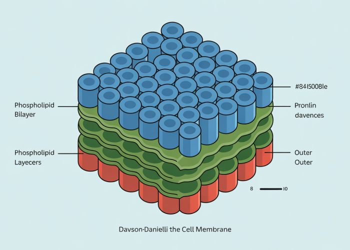
The core concept of the Davson-Danielli model was that the plasma membrane was composed of a lipid bilayer sandwiched between two layers of globular proteins. This "sandwich" structure offered an explanation for several observations prevalent at the time.
The Lipid Bilayer
- Key Component: The central feature was a double layer of phospholipid molecules.
- Arrangement: The hydrophobic tails of the phospholipids faced inward, shielded from the aqueous environment, while the hydrophilic heads faced outward, interacting with water.
- Function: This lipid bilayer was proposed to act as a barrier to the movement of certain substances, especially those that were water-soluble.
The Protein Layers
- Location: The model suggested that layers of proteins coated both the inner and outer surfaces of the lipid bilayer.
- Nature of Proteins: These proteins were assumed to be globular, rather than integral (spanning the membrane).
- Role: These protein layers were thought to provide structural support to the membrane and also to act as pores, allowing the passage of water-soluble molecules that could not cross the lipid bilayer directly.
Evidence Supporting the Davson-Danielli Model
Several lines of evidence initially supported the Davson-Danielli model, leading to its acceptance for several decades.
Chemical Analysis
- Observation: Chemical analysis revealed that plasma membranes contained both lipids and proteins in significant proportions.
- Interpretation: The "sandwich" model neatly accommodated this observation, suggesting a clear physical separation and arrangement of these components.
Surface Tension Measurements
- Observation: The surface tension of cell membranes was found to be much lower than that of pure lipid bilayers.
- Interpretation: Davson and Danielli proposed that the presence of proteins associated with the lipids reduced the surface tension, supporting the idea of protein layers coating the lipid bilayer.
Microscopic Observations
- Observation: Early electron microscopy techniques revealed a trilaminar (three-layered) structure in cell membranes.
- Interpretation: This dark-light-dark appearance was interpreted as the two dark protein layers flanking the lighter lipid bilayer, providing visual confirmation (albeit somewhat misleading in hindsight) of the sandwich model.
The Challenges and Ultimate Rejection of the Model
Despite its initial success, the Davson-Danielli model eventually faced substantial challenges and was ultimately rejected in favor of the Fluid Mosaic Model. Several key pieces of evidence contradicted its fundamental assumptions.
The Problem of Protein Diversity
- Challenge: The model assumed that proteins were uniformly distributed across the membrane surface.
- Contradiction: Research showed that membrane proteins were diverse and had specific functions, suggesting a more complex and heterogeneous arrangement than a simple coating layer.
Freeze-Fracture Electron Microscopy
- Technique: This technique involves freezing a membrane and then fracturing it. The fracture plane often runs along the hydrophobic interior of the lipid bilayer.
- Observation: Freeze-fracture revealed that proteins were embedded within the lipid bilayer, rather than forming distinct surface layers. This contradicted the sandwich model’s depiction of proteins as solely external to the lipid bilayer.
Antibody Staining
- Technique: Antibodies can be used to specifically bind to and label proteins.
- Observation: Antibody staining showed that some membrane proteins were located on only one side of the membrane, while others spanned the entire membrane. This "transmembrane" arrangement was incompatible with the Davson-Danielli model’s two-layer protein structure.
The Hydrophobic Nature of Transmembrane Proteins
- Challenge: The Davson-Danielli model struggled to explain how proteins could exist in the hydrophobic interior of the lipid bilayer.
- Discovery: Researchers discovered that many membrane proteins had hydrophobic regions that allowed them to interact directly with the fatty acid tails of the phospholipids. This contradicted the notion of all proteins being located at the hydrophilic surfaces.
The following table summarizes the main points of contention:
| Feature | Davson-Danielli Model | Observations Leading to Rejection |
|---|---|---|
| Protein Location | Layers coating the surface of the lipid bilayer | Proteins embedded within the lipid bilayer (freeze-fracture) |
| Protein Distribution | Uniformly distributed across the surface | Proteins diverse with specific functions (antibody staining) |
| Protein Transmembrane | No concept of transmembrane proteins | Proteins span the entire membrane (antibody staining, protein analysis) |
| Protein-Lipid Interaction | Indirect; proteins only interacted with lipid head groups | Direct; some proteins have hydrophobic regions interacting with lipid tails |
Davson Danielli Model: Frequently Asked Questions
These FAQs address common questions regarding the Davson Danielli model and its historical context in understanding cell membrane structure.
What exactly was the Davson Danielli model?
The Davson Danielli model, proposed in the 1930s, suggested that the cell membrane was composed of a phospholipid bilayer sandwiched between two layers of protein. Think of it like a sandwich: bread (protein), butter (phospholipids), then more bread (protein).
Why was the Davson Danielli model eventually replaced?
The Davson Danielli model faced challenges because it implied a symmetrical membrane with uniform protein layers. This conflicted with evidence showing the diverse functions and asymmetrical distribution of proteins within the membrane.
What evidence contradicted the Davson Danielli model?
Freeze-fracture electron microscopy revealed that proteins were embedded within the lipid bilayer, not just coating the surfaces as the davson danielli model proposed. Furthermore, biochemical studies showcased the variety of membrane proteins, not just structural ones.
What model replaced the Davson Danielli model?
The Fluid Mosaic Model, proposed by Singer and Nicolson in 1972, superseded the Davson Danielli model. The fluid mosaic model accurately depicts the cell membrane as a dynamic structure with proteins embedded within a fluid phospholipid bilayer, allowing lateral movement.
So, there you have it – a little trip down memory lane with the davson danielli model. While it may not be exactly how we see things now, it certainly paved the way for our current understanding. Pretty cool, right?