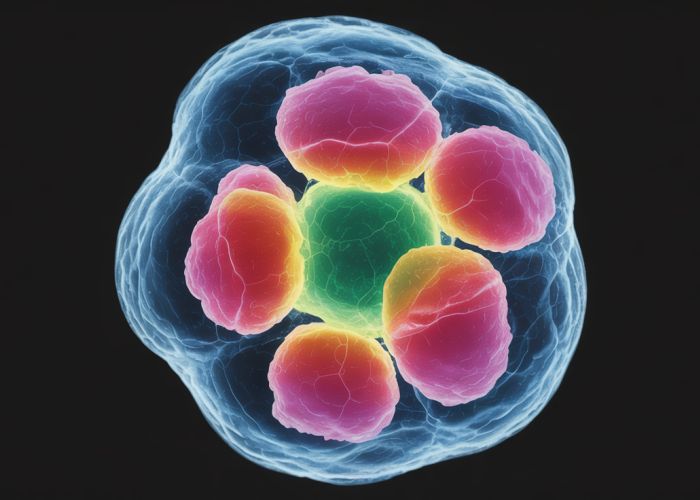
Cytology, a critical branch of biology, studies cell structure, and one key aspect is the color of nucleus. The nucleus, a vital organelle, houses genetic material, and its characteristics are extensively studied in institutions like the National Institutes of Health (NIH). Variations in the color of nucleus can be examined using tools like microscopes, allowing researchers to observe structural details. Scientists such as Rudolf Virchow contributed immensely to understanding cellular processes, laying the foundation for studying the significance of the color of nucleus in cell behavior.
Unlocking the Color of Nucleus: Article Layout Guide
This guide outlines an effective structure for an article focused on understanding the "color of nucleus." The layout is designed to be informative, analytical, and easily digestible for a broad audience.
I. Introduction: Setting the Stage
- Begin with a concise and engaging introduction.
- Briefly define what a nucleus is and its fundamental role within a cell.
- Immediately address the core question: "Does the nucleus have a color?" Avoid ambiguity from the start.
- Present a high-level overview of what the article will cover. Mention that the apparent color is usually due to staining techniques used in observation, not inherent pigmentation.
II. Understanding the Nucleus
- Provide a more detailed explanation of the nucleus and its components.
- Mention the nuclear envelope, chromatin (DNA and proteins), nucleolus, and nuclear pores.
- Briefly touch upon the function of each component.
III. The Concept of "Color" in Microscopy
- Explain why we see color when observing cells under a microscope.
- Describe how microscopes work and how light interacts with biological samples.
- Introduce the concept of staining:
- Explain that staining is a technique used to enhance contrast and visibility.
- Mention that stains bind to specific cellular structures, making them visible.
- Emphasize that the observed "color of nucleus" is generally not its natural color but rather the color of the stain it has absorbed.
IV. Common Staining Techniques and Their Effects on Nuclear Appearance
-
Describe commonly used stains in cell biology, focusing on their effect on nuclear visualization.
- Hematoxylin and Eosin (H&E):
- Explain that hematoxylin stains acidic structures (like DNA in the nucleus) a blue-purple color.
- Describe how eosin stains basic structures (like cytoplasm) pink.
- DAPI (4′,6-diamidino-2-phenylindole):
- Explain that DAPI is a fluorescent stain that binds strongly to DNA.
- Describe how DAPI emits blue light when excited by ultraviolet light.
- Giemsa Stain:
- Explain that Giemsa stain is commonly used to stain blood cells and chromosomes.
- Describe how it can stain the nucleus various shades of purple and pink.
- Hematoxylin and Eosin (H&E):
-
Provide a table summarizing the common stains, their target structures, and resulting colors.
Stain Target Structure Resulting Color Notes Hematoxylin Nucleic Acids (DNA) Blue-Purple Most commonly used stain for nuclear visualization. Eosin Cytoplasm Pink Often used in conjunction with Hematoxylin. DAPI DNA Blue (Fluorescent) Requires UV light excitation; good for visualizing DNA structure. Giemsa Chromosomes, Nuclei Purple, Pink Commonly used in hematology.
V. Natural Pigmentation vs. Staining
- Distinguish between true pigmentation and staining artifacts.
- Explain that some cells do contain natural pigments (e.g., melanin in skin cells).
- State that nuclei generally lack inherent pigmentation that would cause them to have a strong, natural color observable without staining.
- Highlight that even if some components within the nucleus possess some slight color, the overall effect is usually too weak to be noticeable without enhancement techniques.
VI. Factors Affecting the Perceived "Color of Nucleus"
- Discuss other factors besides the stain itself that can influence the observed color.
- Sample Preparation:
- Explain how fixation techniques can alter cellular structures and affect stain binding.
- Mention that dehydration and embedding procedures can also introduce artifacts.
- Microscopy Settings:
- Explain how the microscope’s light source, filters, and camera settings can affect the perceived color.
- Mention that digital image processing can also alter colors.
- Staining Protocols:
- Explain that the concentration of the stain, staining time, and washing steps can all affect the intensity and hue of the color.
- Sample Preparation:
VII. Implications and Applications
- Discuss the importance of understanding staining techniques in various fields.
- Medical Diagnosis: Explain how staining is crucial for identifying cancerous cells, infections, and other diseases by examining the morphology and characteristics of nuclei.
- Research: Explain how staining helps researchers study cell biology, genetics, and developmental biology by visualizing nuclear structures and processes.
- Education: Explain how stained slides are used to teach students about cell structure and function.
VIII. Future Directions
- Briefly mention emerging techniques in cell imaging.
- Label-free Imaging: Discuss methods that allow visualization of cells without staining, such as phase-contrast microscopy or quantitative phase imaging. Explain how these techniques allow a more authentic view of cellular structures.
- Advanced Microscopy Techniques: Mention super-resolution microscopy, which can provide highly detailed images of cellular structures without altering the sample’s natural state.
FAQs: Unlocking the Color of Nucleus
Here are some frequently asked questions to further clarify the fascinating topic of nucleus color.
Why is the nucleus important for understanding cell biology?
The nucleus is the control center of the cell, housing the DNA. Observing the color of the nucleus, or changes in its appearance, can indicate cell health, division stage, or even disease processes. It gives us visual cues to cellular activity.
What factors can affect the color of the nucleus?
Several factors influence the color of the nucleus, including the type of staining used in microscopy, the cell’s stage in its life cycle, and the presence of certain cellular components or proteins within it. Fixation techniques can also impact the final observable color.
How does understanding the color of the nucleus help in disease diagnosis?
Variations in the color of the nucleus, its shape, or size can be indicators of disease states, particularly in cancer diagnosis. For example, irregularly shaped or abnormally stained nuclei are often hallmarks of cancerous cells.
Is the actual nucleus naturally colored inside a living cell?
No, the nucleus itself is largely transparent in living cells without staining. The "color of nucleus" we observe is typically the result of dyes or fluorescent markers that bind to specific structures within the nucleus, allowing visualization under a microscope.
So, that’s a wrap on understanding the color of nucleus! Hopefully, this gives you a better picture of what’s going on inside those tiny cells. If you found this interesting, keep exploring – there’s always something new to learn!