The coagulation cascade, a fundamental process in hemostasis, becomes remarkably understandable through Pathoma‘s concise explanations. This critical biological pathway, responsible for forming blood clots, involves a complex interplay of clotting factors. Understanding these factors and their sequential activation is significantly enhanced by resources like Pathoma, a vital study aid for medical students preparing for exams such as the USMLE. Effectively mastering the coagulation cascade pathoma is a key step in understanding many disease processes.
The coagulation cascade, a series of enzymatic reactions, stands as a cornerstone of hemostasis—the body’s intricate defense against bleeding. Understanding this complex process is not merely an academic exercise. It is absolutely critical for medical professionals, from students to seasoned practitioners, to effectively diagnose and manage a wide range of clinical conditions.
Pathoma, with its concise and visually-driven approach to pathology, offers a valuable framework for demystifying the coagulation cascade. This blog post aims to further simplify and clarify Pathoma’s teachings, providing a practical guide to understanding and applying this essential knowledge.
Why Understanding the Coagulation Cascade is Crucial
The coagulation cascade is far more than a theoretical construct. It is a dynamic process that directly impacts patient care. Disruptions in this cascade can lead to a spectrum of disorders, ranging from life-threatening hemorrhages to thrombotic events that can cause strokes or pulmonary embolisms.
-
Diagnostic Acumen: A thorough understanding of the cascade enables clinicians to accurately interpret laboratory tests such as Prothrombin Time (PT) and Partial Thromboplastin Time (PTT), guiding diagnostic and therapeutic decisions.
-
Treatment Strategies: Many common medications, including anticoagulants like warfarin and heparin, directly target specific components of the coagulation cascade. A solid grasp of the cascade allows for the rational selection and monitoring of these therapies.
-
Clinical Context: Conditions like hemophilia, von Willebrand disease, and disseminated intravascular coagulation (DIC) are all rooted in abnormalities within the coagulation cascade. Understanding the underlying mechanisms allows for targeted and effective management.
Pathoma: A Resource for Learning Pathology
Pathoma, developed by Dr. Hussain Sattar, has become a staple resource for medical students and healthcare professionals seeking a clear and concise understanding of pathology. Its strength lies in its ability to distill complex topics into manageable concepts, often using visual aids and mnemonics to enhance retention.
Pathoma’s coverage of the coagulation cascade is particularly helpful, providing a simplified yet comprehensive overview of the key factors, pathways, and regulatory mechanisms involved. It emphasizes the clinical relevance of each component, connecting basic science principles to real-world patient scenarios.
Our Goal: Simplifying the Cascade with Pathoma’s Approach
This blog post builds upon Pathoma’s framework to provide an even more accessible and practical guide to the coagulation cascade. Our aim is to break down the complex interactions into digestible steps, using clear explanations and illustrative diagrams inspired by Pathoma’s teaching style.
We focus on clarity and application, ensuring that readers not only understand the individual components of the cascade but also how they interact to achieve hemostasis and how disruptions can lead to disease.
By leveraging Pathoma’s simplified approach, we hope to empower medical students and healthcare professionals with a solid foundation in coagulation, enabling them to confidently approach this critical area of medicine.
Understanding the coagulation cascade’s importance sets the stage for delving into its essential components. Before we can appreciate the intricate dance of enzymatic reactions, we must first meet the players: the clotting factors, vitamins, and ions that orchestrate the formation of a stable blood clot.
The Players: Key Factors and Molecules in the Coagulation Cascade
The coagulation cascade is a complex system, but at its heart lie a set of key players. These include a series of proteins known as clotting factors, as well as essential vitamins and ions. Each component plays a specific role, and understanding these roles is critical to grasping the overall process. Pathoma provides an excellent introduction to these factors, and we will expand on that foundation here.
Clotting Factors: A Numbered Cast
The clotting factors are serine proteases (enzymes) that circulate in the blood in an inactive state. Most are synthesized in the liver. They are generally designated by Roman numerals, reflecting the order of their discovery, not their position in the cascade. Key factors include:
-
Factor I (Fibrinogen): A soluble protein that is converted to insoluble fibrin, the structural basis of a blood clot.
-
Factor II (Prothrombin): Converted to thrombin, a critical enzyme that activates other factors and converts fibrinogen to fibrin.
-
Factor III (Tissue Factor): A cell surface receptor that initiates the extrinsic pathway when exposed to blood.
-
Factor IV (Calcium): An ion required for many steps in the coagulation cascade.
-
Factor V: A cofactor that accelerates the activation of prothrombin by Factor Xa.
-
Factor VII: Activates Factor X in the extrinsic pathway.
-
Factor VIII: A cofactor in the intrinsic pathway; deficiency causes hemophilia A.
-
Factor IX: Activates Factor X in the intrinsic pathway; deficiency causes hemophilia B.
-
Factor X: Activates prothrombin to thrombin in the common pathway.
-
Factor XI: Activated by Factor XII in the intrinsic pathway.
-
Factor XII: Initiates the intrinsic pathway through contact activation.
-
Factor XIII: A transglutaminase that cross-links fibrin monomers, strengthening the clot.
Each of these factors must be activated in a precise sequence for the cascade to proceed correctly. Deficiencies or abnormalities in any of these factors can lead to bleeding disorders.
Vitamin K: The Carboxylation Catalyst
Vitamin K is a crucial cofactor for the synthesis of several clotting factors, specifically Factors II (prothrombin), VII, IX, and X. It is essential for the gamma-carboxylation of glutamic acid residues on these factors. This carboxylation allows the factors to bind calcium, which is necessary for their interaction with phospholipid surfaces and subsequent activation in the coagulation cascade.
Warfarin, a commonly used anticoagulant, inhibits vitamin K epoxide reductase, the enzyme responsible for regenerating vitamin K. This inhibition leads to the production of non-functional clotting factors, thereby reducing the blood’s ability to clot.
Calcium: The Bridging Ion
Calcium ions (Factor IV) play a vital role in multiple steps of the coagulation cascade. They act as bridges, binding clotting factors to negatively charged phospholipid surfaces on platelets and endothelial cells. This binding is essential for the assembly of enzyme complexes that drive the cascade. Calcium is particularly important for the activity of Factors II, VII, IX, and X, as their calcium-binding ability is dependent on Vitamin K.
Without sufficient calcium, the coagulation cascade cannot proceed efficiently, and clot formation is impaired.
Fibrinogen and Thrombin: Central Players in Clot Formation
While the numbered clotting factors drive the cascade, two molecules stand out for their central roles: fibrinogen and thrombin. Fibrinogen (Factor I) is a large, soluble protein that is converted into fibrin, an insoluble protein that forms the meshwork of a blood clot.
This conversion is catalyzed by thrombin (Factor IIa), a serine protease that also activates other clotting factors, amplifying the coagulation response. Thrombin’s role extends beyond fibrin formation, as it also activates Factor XIII, which cross-links fibrin monomers, stabilizing the clot.
For a deeper understanding of these crucial elements, refer to Pathoma’s sections on coagulation factors, vitamin K, and the roles of calcium, fibrinogen, and thrombin in clot formation.
Understanding the coagulation cascade’s importance sets the stage for delving into its essential components. Before we can appreciate the intricate dance of enzymatic reactions, we must first meet the players: the clotting factors, vitamins, and ions that orchestrate the formation of a stable blood clot. With the key players now introduced, we can begin to examine the specific pathways that lead to clot formation, starting with the extrinsic pathway.
Extrinsic Pathway: Triggering the Cascade (Pathoma’s Simplified View)
The extrinsic pathway acts as the rapid initiator of the coagulation cascade. It’s the body’s swift response to tissue injury, quickly setting the stage for more robust clot formation. Pathoma effectively simplifies this pathway, highlighting its key components and their interactions. This section will delve into the extrinsic pathway, offering a clear, step-by-step explanation inspired by Pathoma’s approach.
The Pivotal Role of Tissue Factor
Tissue factor (TF), also known as Factor III or thromboplastin, is the linchpin of the extrinsic pathway. It is a transmembrane glycoprotein present on subendothelial cells, such as fibroblasts and smooth muscle cells. Under normal circumstances, TF is sequestered away from circulating blood components.
However, when blood vessels are injured and these subendothelial cells are exposed, TF comes into contact with Factor VII in the plasma. This exposure is the initiating event of the extrinsic pathway. Think of TF as the trigger that starts the entire cascade.
Activation of Factor VII: The First Step
Upon exposure to tissue factor, Factor VII binds to TF to form the Tissue Factor-Factor VIIa complex. Factor VII is a vitamin K-dependent serine protease that circulates in an inactive form.
The binding to TF induces a conformational change in Factor VII, leading to its activation into Factor VIIa. This complex then catalyzes the activation of Factor X, marking a critical juncture in the coagulation cascade.
While Factor VII is the primary binding partner for Tissue Factor, Factor VIIa can also activate additional Factor VII molecules in a positive feedback loop, amplifying the initial signal.
Connecting to the Common Pathway
The Tissue Factor-Factor VIIa complex doesn’t just activate more Factor VII. It also plays a crucial role in activating Factor X, the first step in the common pathway. This is achieved either directly or through the activation of Factor IX from the intrinsic pathway.
Specifically, the TF-VIIa complex activates Factor X to Factor Xa. Factor Xa, along with its cofactor Factor Va, forms the prothrombinase complex, which is essential for converting prothrombin (Factor II) to thrombin (Factor IIa). Thrombin then converts fibrinogen to fibrin, leading to the formation of the clot.
The activation of Factor X by the extrinsic pathway is tightly regulated. Tissue Factor Pathway Inhibitor (TFPI) inhibits the Tissue Factor-Factor VIIa complex to prevent excessive activation of Factor X.
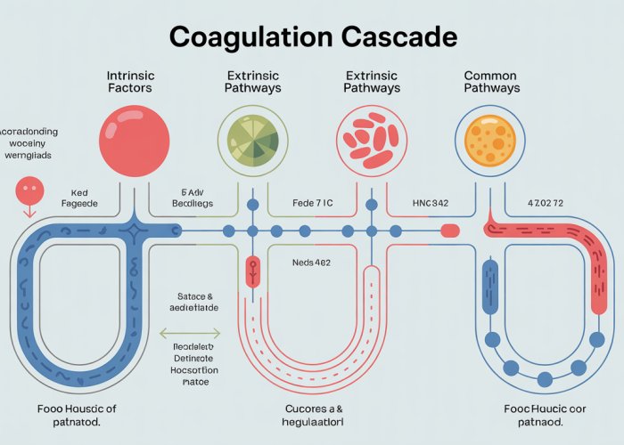
Visualizing the Extrinsic Pathway
A simplified diagram of the extrinsic pathway, inspired by Pathoma, helps to solidify understanding. Imagine a visual representation with the following key elements:
- Tissue factor (TF) displayed on a cell surface.
- Factor VII binding to TF and becoming activated (VIIa).
- The TF-VIIa complex activating Factor X.
- An arrow leading from Factor Xa to the common pathway.
By visualizing these steps, the extrinsic pathway becomes more accessible and easier to remember. The use of visual aids like this one supports Pathoma’s teaching approach.
Intrinsic Pathway: Amplifying the Response (Pathoma’s Approach)
While the extrinsic pathway initiates the clotting process rapidly, the intrinsic pathway acts as a crucial amplifier, generating a sustained and robust response. It’s often referred to as the "contact activation pathway" because it begins when certain plasma proteins come into contact with negatively charged surfaces. Pathoma simplifies the understanding of this pathway, focusing on the key players and their sequential interactions.
The Contact Activation System: Initiating the Intrinsic Pathway
The intrinsic pathway begins with the contact activation system, a complex comprised of Factor XII (Hageman factor), high-molecular-weight kininogen (HMWK), and prekallikrein. Traditionally, it was thought that contact with negatively charged surfaces like glass (in vitro) or collagen (in vivo) was essential for activating Factor XII. However, current understanding suggests that other negatively charged molecules like polyphosphates released from activated platelets may be more physiologically relevant triggers.
Upon contact, prekallikrein is converted to kallikrein by Factor XIIa. Kallikrein then reciprocally activates more Factor XII, creating a positive feedback loop. HMWK acts as a cofactor, facilitating the interaction between prekallikrein and Factor XII.
Factor XII, HMWK, and prekallikrein are not directly involved in in vivo hemostasis, as defects in these factors rarely result in bleeding disorders. Their primary role seems to be more significant in inflammatory and immune responses.
Activation of Factor XI: A Critical Step
Factor XI is activated by Factor XIIa. This activation is crucial for the propagation of the intrinsic pathway. Factor XIa then proceeds to activate Factor IX. Factor XI deficiency (Hemophilia C) is associated with a mild to moderate bleeding tendency, highlighting its importance in the coagulation process.
Factor IX Activation and its Partnership with Factor VIII
Factor IX is activated by Factor XIa in the presence of calcium ions. This activated form, Factor IXa, is essential for the next stage of the intrinsic pathway.
Factor IXa forms a complex with Factor VIIIa, calcium ions, and phospholipid surfaces (typically provided by activated platelets). This complex, known as the tenase complex, is critical for activating Factor X. Factor VIII acts as a cofactor, dramatically enhancing the efficiency of Factor IXa in activating Factor X. Deficiencies in Factor VIII (Hemophilia A) or Factor IX (Hemophilia B) result in significant bleeding disorders, underscoring the importance of the tenase complex.
Bridging to the Common Pathway
The tenase complex (Factor IXa, Factor VIIIa, calcium, and phospholipid) activates Factor X, marking the convergence of the intrinsic and extrinsic pathways onto the common pathway. Once Factor X is activated, the cascade proceeds towards the formation of thrombin and ultimately, the formation of a stable fibrin clot.
By activating Factor X, the intrinsic pathway ensures a sustained production of thrombin, amplifying the initial signal from the extrinsic pathway and leading to robust clot formation. Pathoma emphasizes that a functional intrinsic pathway is therefore essential for effective hemostasis.
The intrinsic and extrinsic pathways, while distinct in their initiation, converge onto a single, unified route: the common pathway. This final stage of the coagulation cascade represents the culmination of all preceding events, ultimately leading to the formation of a stable fibrin clot. Understanding this pathway is crucial, as it’s the point where both the initial triggers of coagulation come together to achieve hemostasis.
Common Pathway: The Final Steps to Clot Formation (Pathoma Explained)
The common pathway is where the intrinsic and extrinsic pathways meet, initiating a sequence of events that results in a stable fibrin clot. This critical stage involves a series of steps, each carefully regulated to ensure efficient and localized clot formation. Pathoma simplifies the understanding of this pathway by focusing on the core components and their interactions.
Role of Factor X and Factor V
Factor X is activated to Factor Xa by both the intrinsic and extrinsic pathways.
Factor Xa then forms a complex with Factor V, calcium ions (Ca2+), and phospholipids on the platelet surface.
Factor V acts as a cofactor, accelerating the activation of prothrombin by Factor Xa. Without Factor V, the activation of prothrombin would be significantly slower and less efficient. This complex is crucial for generating large amounts of thrombin, which is essential for the subsequent steps in the common pathway.
Formation and Function of the Prothrombinase Complex
The complex formed by Factor Xa, Factor Va, calcium, and phospholipids is known as the prothrombinase complex.
This complex is responsible for converting prothrombin (Factor II) to thrombin (Factor IIa).
The prothrombinase complex dramatically enhances the rate of thrombin formation.
The assembly of this complex on the platelet surface ensures that thrombin generation is localized to the site of injury, preventing systemic activation of the coagulation cascade.
Conversion of Prothrombin to Thrombin
Prothrombin, a vitamin K-dependent protein, is cleaved by the prothrombinase complex to form thrombin.
Thrombin is a serine protease that plays a central role in the coagulation cascade.
Thrombin has multiple functions, including:
- Activating more Factors upstream.
- Activating platelets.
- Converting fibrinogen to fibrin.
- Activating Factor XIII.
Its most immediate and crucial role is the conversion of fibrinogen to fibrin, the structural protein of the blood clot.
Conversion of Fibrinogen to Fibrin and the Role of Factor XIII
Fibrinogen, a soluble plasma protein, is converted to fibrin by thrombin. Thrombin cleaves fibrinogen, releasing fibrinopeptides A and B, which allows fibrin monomers to polymerize into a loose fibrin mesh.
This initial fibrin clot is unstable and easily broken down.
Factor XIII, activated by thrombin to Factor XIIIa, then cross-links the fibrin monomers, creating a stable and durable clot. Factor XIIIa forms covalent bonds between glutamine and lysine residues in the fibrin molecules, strengthening the clot and making it resistant to degradation.
The stable fibrin clot provides a framework for wound healing and prevents further blood loss.
Pathoma’s Simplified Explanations
Pathoma emphasizes the importance of understanding the sequential steps in the common pathway. By focusing on the key components – Factor X, Factor V, prothrombin, thrombin, fibrinogen, fibrin, and Factor XIII – Pathoma simplifies the complex interactions, providing a clear and concise overview of this crucial stage in the coagulation cascade.
Pathoma’s visual aids, such as diagrams and flowcharts, further enhance understanding by illustrating the relationships between the different factors and their roles in clot formation.
Regulation of Coagulation: Preventing Excessive Clotting (Pathoma’s Insights)
The coagulation cascade is a powerful and tightly regulated system designed to rapidly respond to vascular injury. However, unchecked activation of this cascade can lead to disastrous consequences, such as thrombosis and disseminated intravascular coagulation (DIC).
Therefore, the body has evolved several elegant mechanisms to prevent excessive clotting and ensure that clot formation remains localized and proportionate to the injury.
Understanding these natural anticoagulant pathways is crucial for comprehending both normal hemostasis and the pathophysiology of various bleeding and thrombotic disorders. Pathoma offers valuable insights into these regulatory mechanisms, simplifying their complexity for medical students and healthcare professionals.
Antithrombin: The Serine Protease Inhibitor
Antithrombin (AT), also known as antithrombin III, is a key player in the regulation of coagulation. It is a serine protease inhibitor (serpin) that primarily inhibits thrombin (Factor IIa) and Factor Xa, but also Factors IXa, XIa, and XIIa to a lesser extent.
Mechanism of Action
Antithrombin functions by forming a stable, irreversible complex with these clotting factors, effectively neutralizing their enzymatic activity. This interaction significantly slows down the coagulation cascade.
Heparin’s Role
The anticoagulant activity of antithrombin is dramatically enhanced by heparin, a naturally occurring glycosaminoglycan. Heparin binds to antithrombin, inducing a conformational change that greatly increases its affinity for its target clotting factors.
This acceleration is clinically exploited in heparin therapy, where heparin is administered to rapidly inhibit coagulation. Heparin essentially acts as a catalyst, speeding up antithrombin’s ability to inactivate thrombin and Factor Xa.
The Protein C and Protein S System: Vitamin K-Dependent Regulators
The Protein C and Protein S system is another crucial anticoagulant pathway that relies on vitamin K-dependent proteins.
Protein C, once activated, inactivates Factors Va and VIIIa, two essential cofactors in the coagulation cascade. Protein S acts as a cofactor for Protein C, enhancing its activity.
Activation of Protein C
Protein C is activated by thrombin bound to thrombomodulin, a receptor on endothelial cells. When thrombin binds to thrombomodulin, its procoagulant activity is switched to an anticoagulant activity. This complex then activates Protein C.
Mechanism of Action
Activated Protein C (APC), with Protein S as its cofactor, degrades Factors Va and VIIIa. Factor V Leiden, a genetic mutation that makes Factor Va resistant to inactivation by APC, is a common cause of inherited thrombophilia.
Tissue Factor Pathway Inhibitor (TFPI): Limiting the Extrinsic Pathway
Tissue Factor Pathway Inhibitor (TFPI) is a protein that inhibits the extrinsic pathway of coagulation. It is synthesized by endothelial cells and inhibits Factor Xa directly.
Mechanism of Action
TFPI forms a complex with Factor Xa, which then inhibits the Tissue Factor-Factor VIIa complex. This effectively shuts down the initiation of coagulation via the extrinsic pathway.
TFPI plays a crucial role in limiting the extent of coagulation following tissue injury and prevents systemic activation of the coagulation cascade.
Pathoma’s Perspective
Pathoma emphasizes that these regulatory mechanisms are essential for maintaining hemostatic balance. Disruptions in these pathways, whether due to genetic deficiencies, acquired conditions, or medications, can lead to either bleeding or thrombotic disorders.
Understanding how these natural anticoagulants work is vital for interpreting laboratory tests, understanding the mechanisms of anticoagulant drugs, and managing patients with coagulation disorders.
By simplifying these complex processes, Pathoma provides a strong foundation for mastering the intricacies of coagulation regulation.
Antithrombin’s critical role in preventing runaway coagulation makes disruptions to its function, or that of other regulatory proteins, clinically significant. These disruptions often manifest as thrombotic disorders, highlighting the delicate balance within the hemostatic system. Now, let’s explore some specific clinical conditions intricately linked to the coagulation cascade, offering a deeper understanding of how breakdowns in this system can lead to disease.
Clinical Relevance: Coagulation Cascade Disorders (Pathoma Connections)
The coagulation cascade isn’t merely an academic exercise; its intricacies directly translate to a range of clinical disorders that profoundly impact patient health. Understanding these connections, particularly as emphasized by Pathoma, is essential for accurate diagnosis and effective management.
Let’s delve into some key clinical conditions that illustrate the practical implications of disruptions within the coagulation cascade:
Hemophilia A and B: Deficiencies in Factors VIII and IX
Hemophilia A and B are classic examples of bleeding disorders caused by deficiencies in crucial clotting factors. Hemophilia A is due to a deficiency in Factor VIII, while Hemophilia B (also known as Christmas disease) results from a deficiency in Factor IX.
Both are X-linked recessive disorders, meaning they predominantly affect males, as they only have one X chromosome. Females, with two X chromosomes, are typically carriers, potentially passing the affected gene to their offspring.
The severity of hemophilia depends on the degree of factor deficiency. Individuals with severe hemophilia experience spontaneous bleeding into joints (hemarthrosis) and muscles, while those with milder forms may only bleed excessively after trauma or surgery.
Pathoma emphasizes the importance of recognizing the clinical presentation of hemophilia, including the characteristic hemarthrosis and prolonged PTT (partial thromboplastin time), while PT (prothrombin time) remains normal. Treatment involves replacement therapy with recombinant Factor VIII or IX, respectively, to restore clotting ability.
Von Willebrand Disease: A Qualitative or Quantitative Defect
Von Willebrand disease (vWD) is the most common inherited bleeding disorder, affecting approximately 1% of the population. Unlike hemophilia, vWD can affect both males and females. It is characterized by a defect in von Willebrand factor (vWF), a protein that plays a crucial role in platelet adhesion and acts as a carrier protein for Factor VIII.
vWF has two primary functions: it mediates the adhesion of platelets to damaged endothelium at sites of vascular injury, and it binds to Factor VIII, protecting it from degradation and delivering it to the site of injury.
There are several types of vWD, classified based on the nature and severity of the vWF defect. Type 1 is the most common and is characterized by a quantitative deficiency of vWF. Type 2 involves qualitative defects in vWF function. Type 3 is the rarest and most severe form, characterized by a near-complete absence of vWF.
Clinical manifestations of vWD include mucocutaneous bleeding, such as nosebleeds, easy bruising, and prolonged bleeding after surgery or dental procedures. In women, menorrhagia (heavy menstrual bleeding) is a common symptom.
Pathoma highlights the importance of differentiating vWD from other bleeding disorders, emphasizing the characteristic prolonged bleeding time and abnormal vWF assays. Treatment options include desmopressin (DDAVP), which stimulates the release of vWF from endothelial cells, and vWF concentrates derived from plasma.
Factor V Leiden: Hypercoagulability and Thrombosis
Factor V Leiden is the most common inherited hypercoagulability disorder, affecting approximately 5% of the Caucasian population. It is caused by a specific mutation in the Factor V gene, resulting in a Factor V protein that is resistant to inactivation by activated protein C (APC).
The protein C pathway is a crucial anticoagulant mechanism that normally inactivates Factors Va and VIIIa, thereby limiting the coagulation cascade. In individuals with Factor V Leiden, APC is unable to effectively inactivate Factor V, leading to a prolonged procoagulant state.
This increased risk of thrombosis primarily manifests as deep vein thrombosis (DVT) and pulmonary embolism (PE), particularly in the context of other risk factors such as surgery, pregnancy, or oral contraceptive use.
Pathoma emphasizes the importance of considering Factor V Leiden in patients presenting with unexplained venous thromboembolism, especially at a young age or with a family history of thrombosis. Diagnosis is typically made through genetic testing.
Disseminated Intravascular Coagulation (DIC): A Complex, Life-Threatening Condition
Disseminated intravascular coagulation (DIC) is a complex and life-threatening condition characterized by widespread activation of the coagulation cascade, leading to simultaneous thrombosis and hemorrhage. It is not a primary disease but rather a secondary complication of other underlying conditions, such as sepsis, trauma, malignancy, and obstetric complications.
In DIC, uncontrolled activation of the coagulation cascade results in the formation of numerous microthrombi throughout the vasculature, leading to organ ischemia and damage. Simultaneously, the consumption of clotting factors and platelets leads to a bleeding diathesis, resulting in widespread hemorrhage.
Pathoma stresses that DIC is a consumptive coagulopathy, meaning that the body’s clotting resources are being used up faster than they can be replenished. This leads to a paradoxical situation where the patient is both clotting and bleeding at the same time.
Clinical manifestations of DIC include widespread petechiae, purpura, and ecchymoses, as well as bleeding from puncture sites, mucous membranes, and surgical wounds. Organ dysfunction due to microthrombi can also occur, leading to acute respiratory distress syndrome (ARDS), renal failure, and neurological complications.
Laboratory findings in DIC typically include prolonged PT and PTT, decreased platelet count, elevated D-dimer levels (indicating fibrinolysis), and decreased fibrinogen levels. Treatment of DIC focuses on addressing the underlying cause and providing supportive care, including blood transfusions and clotting factor replacement.
Pathoma provides a clear framework for understanding the pathogenesis, clinical presentation, and laboratory findings of DIC, highlighting its importance as a medical emergency requiring prompt diagnosis and treatment.
Clinical disorders stemming from coagulation cascade abnormalities vividly illustrate the system’s importance. From the bleeding diatheses of hemophilia to the hypercoagulable state of Factor V Leiden, disruptions in the cascade have significant implications. But identifying these disruptions requires more than just clinical suspicion. Laboratory tests play a crucial role in diagnosing and monitoring coagulation disorders, offering a window into the intricate workings of the cascade itself.
Laboratory Tests: Assessing the Coagulation Cascade (Pathoma’s Perspective)
Two cornerstone laboratory tests, Prothrombin Time (PT) and Partial Thromboplastin Time (PTT), are indispensable tools for evaluating the coagulation cascade. These assays provide valuable insights into the functionality of different pathways within the cascade, aiding in the diagnosis of bleeding disorders, monitoring anticoagulant therapy, and assessing overall coagulation status. Pathoma emphasizes the importance of understanding the principles behind these tests and their clinical interpretation.
Prothrombin Time (PT): Evaluating the Extrinsic and Common Pathways
The Prothrombin Time (PT) is a laboratory test that assesses the integrity of the extrinsic and common pathways of the coagulation cascade. This test measures the time it takes for a clot to form in a plasma sample after the addition of thromboplastin (tissue factor) and calcium.
The PT is primarily sensitive to deficiencies or inhibitors of factors in the extrinsic pathway, specifically Factor VII, as well as factors in the common pathway (Factors X, V, Prothrombin [II], and Fibrinogen [I]).
Factors Involved in PT
- Factor VII: The key initiator of the extrinsic pathway.
- Factor X: Activated by Factor VIIa in conjunction with tissue factor.
- Factor V: A cofactor in the prothrombinase complex.
- Prothrombin (Factor II): The precursor to thrombin.
- Fibrinogen (Factor I): Converted to fibrin, forming the clot matrix.
An prolonged PT indicates a deficiency or inhibition of one or more of these factors, which can be caused by various conditions such as vitamin K deficiency, liver disease, warfarin therapy, or factor deficiencies.
Partial Thromboplastin Time (PTT): Assessing the Intrinsic and Common Pathways
The Partial Thromboplastin Time (PTT) evaluates the intrinsic and common pathways of the coagulation cascade. In this test, plasma is activated with a contact activator (e.g., kaolin, silica) and phospholipids, followed by the addition of calcium. The time it takes for a clot to form is then measured.
The PTT is sensitive to deficiencies or inhibitors of factors in the intrinsic pathway (Factors XII, XI, IX, VIII) and the common pathway (Factors X, V, Prothrombin [II], and Fibrinogen [I]).
Factors Involved in PTT
- Factor XII, HMWK, Prekallikrein: Components of the contact activation system.
- Factor XI: Activated by Factor XIIa.
- Factor IX: Activated by Factor XIa; crucial for activating Factor X in the intrinsic pathway.
- Factor VIII: A cofactor for Factor IXa.
- Factor X: Activated by Factor IXa and Factor VIIIa.
- Factor V: A cofactor in the prothrombinase complex.
- Prothrombin (Factor II): The precursor to thrombin.
- Fibrinogen (Factor I): Converted to fibrin, forming the clot matrix.
A prolonged PTT suggests a deficiency or inhibition of one or more of these factors, as seen in conditions like hemophilia (Factor VIII or IX deficiency), lupus anticoagulants, heparin therapy, or other factor deficiencies.
Monitoring Anticoagulation Therapy with PT and PTT
PT and PTT are also critical for monitoring the effectiveness of anticoagulant medications. The choice of test depends on the specific anticoagulant being used.
-
Warfarin: Warfarin inhibits the vitamin K-dependent synthesis of Factors II, VII, IX, and X. Since Factor VII has the shortest half-life, the PT is particularly sensitive to warfarin’s effects and is used to monitor warfarin therapy, typically reported as an INR (International Normalized Ratio) to standardize results.
-
Heparin: Heparin enhances the activity of antithrombin, which inhibits thrombin and Factor Xa. The PTT is used to monitor unfractionated heparin therapy. Low-molecular-weight heparin (LMWH) has a more predictable effect and often does not require routine PTT monitoring.
Pathoma’s Perspective on PT and PTT
Pathoma highlights the clinical significance of PT and PTT in diagnosing bleeding disorders and monitoring anticoagulation. Pathoma also underscores the importance of understanding which factors are assessed by each test. By correlating PT and PTT results with clinical findings, one can pinpoint specific factor deficiencies or inhibitors, guiding appropriate management strategies. Pathoma presents clear diagrams and explanations that are invaluable for understanding these tests and their interpretations.
Clinical disorders stemming from coagulation cascade abnormalities vividly illustrate the system’s importance. From the bleeding diatheses of hemophilia to the hypercoagulable state of Factor V Leiden, disruptions in the cascade have significant implications. But identifying these disruptions requires more than just clinical suspicion. Laboratory tests play a crucial role in diagnosing and monitoring coagulation disorders, offering a window into the intricate workings of the cascade itself.
Now, let’s move beyond diagnosis and consider how we can therapeutically intervene in the coagulation cascade. A range of anticoagulant medications target specific points within this intricate system, offering crucial tools for managing and preventing thromboembolic events. Understanding their mechanisms of action is essential for effective and safe clinical practice.
Anticoagulant Medications: Targeting the Coagulation Cascade
Anticoagulant medications are vital in preventing and treating thromboembolic diseases. These drugs work by interfering with the coagulation cascade at various points, ultimately reducing the formation of blood clots.
A deep understanding of these medications—their mechanisms, indications, and potential adverse effects—is critical for all healthcare professionals. Let’s explore some of the most commonly used anticoagulants and their specific targets within the coagulation cascade.
Warfarin: Inhibiting Vitamin K Epoxide Reductase
Warfarin remains a widely used anticoagulant, particularly for long-term management of thromboembolic conditions. Its mechanism of action centers on disrupting the vitamin K-dependent synthesis of several clotting factors.
Specifically, warfarin inhibits the vitamin K epoxide reductase (VKORC1) enzyme. This enzyme is essential for regenerating the active form of vitamin K, which is required for the carboxylation of factors II, VII, IX, and X, as well as proteins C and S.
By inhibiting VKORC1, warfarin effectively reduces the levels of these functional clotting factors. This leads to a decreased ability of the blood to clot.
The effect of warfarin is not immediate because it affects the synthesis of new clotting factors. It does not directly inactivate existing factors. This delayed onset is crucial to understand when initiating warfarin therapy.
Regular monitoring of the International Normalized Ratio (INR) is essential to ensure the drug remains within the therapeutic range. This is because the drug has a narrow therapeutic window.
Heparin: Activating Antithrombin
Heparin is another crucial anticoagulant, often used in acute settings due to its rapid onset of action. Unlike warfarin, heparin does not directly inhibit clotting factors. Instead, it acts as a catalyst to accelerate the activity of antithrombin (AT).
Antithrombin is a naturally occurring inhibitor of several coagulation factors, including thrombin (Factor IIa) and Factor Xa. Heparin binds to antithrombin, causing a conformational change that greatly enhances its ability to inhibit these factors.
There are different types of heparin, including unfractionated heparin (UFH) and low-molecular-weight heparin (LMWH).
-
Unfractionated Heparin (UFH): UFH is a mixture of polysaccharide chains of varying lengths. It effectively inhibits both thrombin and Factor Xa, but requires close monitoring of the activated partial thromboplastin time (aPTT).
-
Low-Molecular-Weight Heparin (LMWH): LMWHs, such as enoxaparin and dalteparin, have a more predictable response and primarily inhibit Factor Xa. They typically do not require routine aPTT monitoring.
Protamine sulfate is used to reverse the effects of heparin in case of excessive bleeding. This is very important.
Direct Thrombin Inhibitors (DTIs)
Direct Thrombin Inhibitors (DTIs) represent a class of anticoagulants that directly bind to and inhibit thrombin (Factor IIa). This is regardless of whether thrombin is free in the circulation or bound to fibrin in a clot.
Unlike heparin, DTIs do not require antithrombin as a cofactor. This makes them effective in patients with antithrombin deficiency. Examples of DTIs include dabigatran, argatroban, and bivalirudin.
-
Dabigatran: An oral DTI that directly inhibits both free and clot-bound thrombin.
-
Argatroban and Bivalirudin: Typically administered intravenously, often used in patients with heparin-induced thrombocytopenia (HIT).
DTIs offer a more predictable anticoagulant effect compared to heparin. However, specific reversal agents, such as idarucizumab (for dabigatran), are available in case of bleeding complications.
Direct Factor Xa Inhibitors
Direct Factor Xa inhibitors are another class of oral anticoagulants that selectively inhibit Factor Xa, a key enzyme in the common pathway of the coagulation cascade. By inhibiting Factor Xa, these drugs prevent the conversion of prothrombin to thrombin, thereby reducing clot formation.
Commonly used Factor Xa inhibitors include rivaroxaban, apixaban, edoxaban, and betrixaban.
These medications offer several advantages, including oral administration, fixed dosing, and no routine monitoring.
Similar to DTIs, specific reversal agents, such as andexanet alfa, are available for some Factor Xa inhibitors in case of major bleeding events.
Connecting Medications to the Cascade Diagram
Visualizing where these medications act within the coagulation cascade is crucial for understanding their mechanisms.
-
Warfarin: Acts upstream by reducing the synthesis of multiple vitamin K-dependent clotting factors (II, VII, IX, X).
-
Heparin: Enhances the activity of antithrombin, which inhibits thrombin (IIa) and Factor Xa.
-
Direct Thrombin Inhibitors: Directly inhibit thrombin (IIa), preventing its role in converting fibrinogen to fibrin.
-
Direct Factor Xa Inhibitors: Directly inhibit Factor Xa, preventing the formation of thrombin.
By understanding these relationships, healthcare professionals can better appreciate the nuances of anticoagulant therapy and make informed decisions regarding drug selection and management.
Coagulation Cascade: Pathoma Simplified! – FAQs
Here are some frequently asked questions to further clarify the coagulation cascade, as presented in Pathoma. Hopefully, these answers help solidify your understanding!
What’s the most important takeaway from the Pathoma approach to the coagulation cascade?
Pathoma emphasizes understanding the why behind each step. Instead of rote memorization, focus on the roles of factors, the points of amplification, and how deficiencies lead to specific bleeding disorders. Understanding the big picture is key to mastering the coagulation cascade pathoma.
How does Pathoma help simplify remembering the intrinsic and extrinsic pathways?
Pathoma focuses on key initiating events for each pathway. For the intrinsic pathway, remember the "subendothelial collagen" trigger. The extrinsic pathway is triggered by "tissue thromboplastin." This simplified framework makes remembering the coagulation cascade pathoma much easier.
What is the significance of Vitamin K in the coagulation cascade pathoma explained?
Vitamin K is crucial for the gamma-carboxylation of several coagulation factors (II, VII, IX, and X). This modification is essential for their calcium-binding ability, which is vital for their function in the coagulation cascade. Pathoma stresses its importance as a target for anticoagulants like Warfarin.
How do Pathoma’s explanations connect the coagulation cascade to clinical scenarios?
Pathoma consistently connects the coagulation cascade to real-world diseases like hemophilia (factor VIII or IX deficiency) or disseminated intravascular coagulation (DIC). By understanding the underlying mechanism of the coagulation cascade pathoma, you can better predict and understand these clinical presentations.
So, that’s the gist of coagulation cascade pathoma! Hopefully, this helped untangle things a bit. Now go forth and conquer those medical mysteries!