The neuron, a fundamental unit of the nervous system, relies heavily on the integrity of its cell body neuron, also known as the soma. Neurotransmitters, key chemical messengers, are synthesized within this crucial location. Understanding the structure of the cell body neuron is paramount for researchers at institutions like the National Institutes of Health (NIH), who investigate neuronal function and disease. The health of the cell body neuron ultimately influences its electrical signal conduction, observable through techniques like electrophysiology.
The nervous system, a vast and intricate network, governs every aspect of our being, from the simplest reflexes to the most complex thoughts and emotions. At the heart of this system lie neurons, specialized cells responsible for transmitting information throughout the body. Understanding the structure and function of these remarkable cells is paramount to unraveling the mysteries of the brain and nervous system.
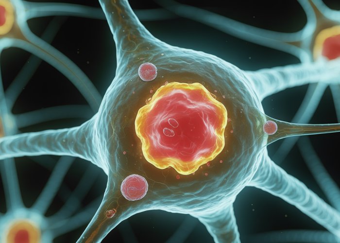
The Neuron: The Nervous System’s Building Block
The neuron is the fundamental unit of the nervous system, a highly specialized cell designed for rapid communication. These cells form complex circuits and networks, enabling us to perceive the world, make decisions, and execute actions.
Like all cells, neurons possess a cell body, or soma, which serves as the neuron’s control center.
The Soma: The Neuron’s Command Center
The soma is the central hub of the neuron, housing the nucleus and other essential organelles. It plays a critical role in maintaining the neuron’s health, synthesizing proteins, and integrating incoming signals.
Think of the soma as the neuron’s executive office, where crucial decisions are made and vital functions are coordinated.
Exploring the Intricacies of the Soma: A Thesis
This article will delve into the intricate details of the soma, exploring its structure, function, and significance in neuronal communication. We will examine the various components of the soma, from the nucleus to the cytoplasm, and discuss their respective roles in neuronal activity.
Furthermore, we will explore how the soma integrates incoming signals, generates action potentials, and contributes to the synthesis of essential proteins. By understanding the soma, we can gain valuable insights into the complex workings of the nervous system and its impact on our overall health and well-being.
The preceding discussion has established the soma as the neuron’s control center, where critical decisions are made and vital functions are coordinated. Now, let us embark on a detailed exploration of the soma’s internal architecture, examining the key components that enable it to perform its essential roles.
Anatomy of the Soma: A Deep Dive into its Components
The soma, also known as the cell body, is the central part of a neuron that contains the nucleus and other vital organelles. It’s typically located between the dendrites, which receive incoming signals, and the axon, which transmits signals to other neurons or cells.
Defining the Soma: Location and General Structure
The soma serves as the neuron’s command center, integrating incoming signals and coordinating metabolic activities. It’s the primary site for protein synthesis and other essential cellular processes necessary for neuronal function. Its shape can vary widely depending on the type of neuron.
The soma is usually more or less spherical or pyramidal. It is defined by the lipid bilayer, which isolates the inner cell organelles from the extracellular environment.
The Nucleus: The Neuron’s Central Command
At the heart of the soma lies the nucleus, the neuron’s control center. The nucleus houses the neuron’s genetic material in the form of DNA, organized into chromosomes. It is the blueprint that dictates the cell’s structure, function, and development.
Directing Cellular Activities and Gene Expression
The nucleus plays a critical role in directing cellular activities by regulating gene expression. Through transcription and translation, the nucleus controls the synthesis of proteins, which are the workhorses of the cell. These proteins are essential for maintaining the neuron’s structure, function, and communication with other cells.
The Cytoplasm: The Intracellular Environment
The cytoplasm is the gel-like substance that fills the soma, surrounding the nucleus and other organelles.
It’s primarily composed of water, ions, enzymes, and other molecules that are essential for cellular function.
Suspension of Organelles and Transport of Substances
The cytoplasm serves as a medium for suspending organelles, allowing them to move freely and interact with each other. It also facilitates the transport of substances within the soma, including nutrients, metabolites, and signaling molecules. This transport is crucial for maintaining cellular homeostasis and supporting neuronal activity.
Key Organelles within the Soma: A Functional Overview
The cytoplasm is home to a variety of organelles, each with its specific function. These organelles work together to maintain the neuron’s health, synthesize proteins, and generate energy.
- Endoplasmic Reticulum (ER): A network of interconnected membranes involved in protein synthesis, folding, and lipid metabolism. The rough ER, studded with ribosomes, is primarily responsible for protein synthesis, while the smooth ER plays a role in lipid metabolism and detoxification.
- Golgi Apparatus: This organelle processes and packages proteins synthesized in the ER. It modifies proteins by adding carbohydrates or lipids and then sorts them into vesicles for transport to other parts of the neuron or for secretion.
- Mitochondria: Often referred to as the "powerhouse" of the cell, mitochondria generate energy in the form of ATP through cellular respiration. Neurons have high energy demands, making mitochondria particularly abundant within the soma.
- Ribosomes: These are the sites of protein synthesis. They can be found free-floating in the cytoplasm or attached to the rough ER. Ribosomes translate mRNA into proteins, using the genetic code as a guide.
Cell Membrane: The Gatekeeper
The cell membrane, also known as the plasma membrane, is the outer boundary of the soma, enclosing its contents and separating them from the external environment.
It’s composed of a lipid bilayer with embedded proteins.
The lipid bilayer provides a barrier that prevents the free passage of molecules, while the embedded proteins regulate the transport of specific substances across the membrane. This selective permeability is crucial for maintaining cell integrity and regulating the flow of ions necessary for action potential generation.
The preceding discussion has established the soma as the neuron’s control center, where critical decisions are made and vital functions are coordinated. Now, let us embark on a detailed exploration of the soma’s internal architecture, examining the key components that enable it to perform its essential roles.
Functional Significance: The Soma’s Multifaceted Roles in Neuronal Activity
The soma, far from being a mere housing for the nucleus, is a dynamic hub of neuronal activity. It’s the neuron’s central processing unit, orchestrating a multitude of functions vital for communication and survival.
These functions include the integration of incoming signals, the initiation of action potentials, the synthesis of proteins, and the production of neurotransmitters. Understanding these multifaceted roles is crucial for comprehending the complex operations of the nervous system.
Integration of Signals: The Soma as Decision-Maker
The neuron is constantly bombarded with signals from other neurons. These signals arrive at the dendrites and are then transmitted to the soma. The soma acts as a sophisticated integrator, processing these incoming signals to determine whether or not to fire an action potential.
This integration process is not a simple summation; it’s a complex calculation involving both the strength and timing of the incoming signals. Two key mechanisms govern this integration: spatial and temporal summation.
Spatial Summation
Spatial summation refers to the integration of signals arriving from different locations on the neuron simultaneously. If multiple signals arrive at the soma at the same time, their effects are added together.
If the combined effect is strong enough, it can trigger an action potential. This allows the neuron to respond to a coordinated input from multiple sources.
Temporal Summation
Temporal summation, on the other hand, involves the integration of signals arriving at the soma at slightly different times. If a signal arrives at the soma and is quickly followed by another signal from the same source, their effects can be added together.
This happens because the first signal has not yet completely dissipated when the second signal arrives. Temporal summation allows the neuron to respond to a rapid sequence of inputs from a single source.
Generation of Action Potential: Initiating the Nerve Impulse
If the integrated signal at the soma reaches a certain threshold, it triggers the generation of an action potential. The action potential is a rapid change in electrical potential across the neuron’s membrane that travels down the axon.
The axon hillock, a specialized region of the soma where the axon originates, plays a crucial role in initiating the action potential. It has a high concentration of voltage-gated sodium channels. These channels open when the membrane potential reaches the threshold, causing a rapid influx of sodium ions into the cell.
This influx of sodium ions causes the membrane potential to rapidly depolarize, initiating the action potential.
Threshold and Depolarization
The threshold is the minimum level of depolarization required to trigger an action potential. Once the threshold is reached, the action potential is generated in an all-or-nothing fashion.
Depolarization refers to the decrease in the potential difference across the neuron’s membrane, making the inside of the cell less negative relative to the outside. The rapid influx of sodium ions during depolarization causes the membrane potential to swing positive.
Protein Synthesis: Building Blocks of Neuronal Function
The soma is the primary site of protein synthesis in the neuron. Proteins are essential for virtually every aspect of neuronal function, including:
- Enzymes: Catalyze biochemical reactions.
- Receptors: Bind to neurotransmitters and other signaling molecules.
- Structural proteins: Maintain the shape and structure of the neuron.
- Ion channels: Regulate the flow of ions across the cell membrane.
The nucleus, located within the soma, contains the genetic information (DNA) required for protein synthesis. This information is transcribed into messenger RNA (mRNA), which is then transported to the ribosomes in the cytoplasm. The ribosomes then translate the mRNA into proteins.
Neurotransmitter Synthesis: Chemical Messengers of the Brain
In addition to protein synthesis, the soma is also involved in the synthesis of neurotransmitters. Neurotransmitters are chemical messengers that transmit signals from one neuron to another across a synapse.
The enzymes required for neurotransmitter synthesis are produced in the soma and then transported to the axon terminals, where neurotransmitters are synthesized and packaged into vesicles. Different types of neurons synthesize different types of neurotransmitters.
This allows for a wide range of signaling possibilities in the nervous system. The soma’s involvement in neurotransmitter production highlights its crucial role in neuronal communication.
The soma’s multifaceted functions, including signal integration, action potential generation, and protein synthesis, are all intrinsically linked to its ability to communicate effectively with other neuronal structures. It’s a bidirectional conversation; the soma not only receives information but also transmits it, playing a critical role in the overall flow of information within the nervous system.
Communication Pathways: The Soma’s Connections to Other Neuronal Structures
The neuron, in its essence, is a communication specialist. The soma, as its central processing unit, is strategically connected to other parts of the neuron, allowing for seamless communication and signal transmission. Understanding these connections is vital to appreciating the neuron’s overall function within the complex network of the nervous system.
Dendritic Connections: Receiving the Message
Dendrites are the neuron’s antenna, branching out to receive signals from countless other neurons. These signals, often in the form of neurotransmitters, bind to receptors on the dendrites, initiating a cascade of electrical and chemical events.
The dendrites then act as messengers, transmitting these signals to the soma for processing.
The Structure of Dendrites and Synaptic Connections
The intricate, tree-like structure of dendrites is no accident. This morphology dramatically increases the surface area available for synaptic connections, allowing a single neuron to receive input from thousands of other neurons.
These connections, called synapses, are the points of contact between neurons where signals are transmitted. The density and distribution of synapses on the dendrites directly influence the neuron’s ability to integrate and respond to incoming information.
The Axon and Axon Hillock: Sending the Response
Once the soma has integrated the incoming signals and "decided" to fire an action potential, it’s the axon’s job to carry that signal to other neurons, muscles, or glands. The axon is the neuron’s output pathway, a long, slender projection that extends from the soma.
The Axon Hillock: Where the Signal Begins
The axon hillock is a specialized region of the soma where the axon originates. This area is crucial for initiating action potentials because it has a high concentration of voltage-gated sodium channels. These channels are essential for the rapid influx of sodium ions that drives the depolarization phase of the action potential.
The axon hillock acts as a gatekeeper, ensuring that the signal reaching the axon is strong enough to trigger an action potential.
The Cell Membrane: Maintaining Integrity and Regulating Flow
The cell membrane, also known as the plasma membrane, is a vital structure that surrounds the soma and all other parts of the neuron. It’s composed of a lipid bilayer with embedded proteins, acting as a selective barrier. This barrier both maintains the integrity of the cell and regulates the flow of ions and molecules in and out.
This regulation is essential for maintaining the resting membrane potential, a crucial factor in the neuron’s ability to generate and transmit action potentials. Ion channels within the cell membrane allow specific ions, such as sodium, potassium, and chloride, to cross the membrane under controlled conditions, driving the electrical events underlying neuronal communication.
The Soma’s Role in Spinal Cord and Brain Communication
The soma is not an isolated entity; it’s an integral part of the vast communication network within the central nervous system. Somata are strategically positioned throughout the brain and spinal cord to receive, process, and transmit signals effectively.
In the spinal cord, somata of motor neurons reside in the ventral horn, receiving input from interneurons and upper motor neurons to control muscle movements.
In the brain, somata are found in various regions, from the cerebral cortex to the deep brain nuclei, where they participate in complex neural circuits underlying everything from sensory perception to cognition.
The Soma and the Central Nervous System (CNS)
The soma’s involvement in communication and transmission within the CNS is all-encompassing. It’s the point of convergence for signals from the periphery, the site of integration for complex computations, and the origin of outgoing signals that control various bodily functions.
The soma’s role extends beyond simply relaying information. It actively participates in shaping the information flow within the CNS, influencing neural plasticity and contributing to learning and memory. Disruptions in soma function can have profound consequences for the entire nervous system, leading to a wide range of neurological disorders.
The exchange of information between neurons is paramount, but the neuron doesn’t operate in isolation. It exists within a complex milieu of other cells, particularly glial cells, which play indispensable supportive roles. Their influence extends to virtually every aspect of neuronal function, making them critical partners in the symphony of the nervous system.
Supporting Cast: The Crucial Role of Glial Cells in Soma Function
While neurons rightly receive much of the spotlight, the importance of glial cells in supporting neuronal function cannot be overstated. These cells, often called neuroglia, outnumber neurons in the brain and perform a diverse array of essential tasks.
They are not merely passive bystanders; instead, they actively contribute to the health, maintenance, and efficient operation of neurons, including the soma. This section will explore the major types of glial cells and their specific interactions with the soma, highlighting their vital contribution to neuronal well-being.
Glial Cells: More Than Just Glue
The term "glia" is derived from the Greek word for "glue," reflecting the historical belief that these cells simply held neurons together. However, modern neuroscience has revealed that glial cells are far more than mere structural support.
They are active participants in neuronal communication, providing essential nutrients, regulating the extracellular environment, and even influencing synaptic transmission. Understanding their multifaceted roles is crucial for a complete picture of neuronal function.
Types of Glial Cells and Their Functions
There are several distinct types of glial cells in the nervous system, each with specialized functions:
- Astrocytes: These star-shaped cells are the most abundant glial cells in the brain and spinal cord. They perform a wide range of functions, including:
- Providing structural support to neurons and blood vessels.
- Maintaining the blood-brain barrier, which protects the brain from harmful substances.
- Regulating the chemical environment around neurons by absorbing excess neurotransmitters and ions.
- Supplying nutrients to neurons.
- Oligodendrocytes: These cells are responsible for myelination in the central nervous system (brain and spinal cord).
- Myelin is a fatty substance that insulates axons, allowing for faster and more efficient signal transmission.
- Each oligodendrocyte can myelinate multiple axons.
- Schwann Cells: These cells perform a similar function to oligodendrocytes, but in the peripheral nervous system (nerves outside the brain and spinal cord).
- Each Schwann cell myelinates only one segment of a single axon.
- Microglia: These are the resident immune cells of the brain and spinal cord.
- They act as scavengers, removing debris, dead cells, and pathogens.
- Microglia also play a role in synaptic pruning and remodeling.
- Ependymal Cells: These cells line the ventricles of the brain and the central canal of the spinal cord.
- They produce cerebrospinal fluid (CSF), which cushions and protects the brain and spinal cord.
- Ependymal cells also have cilia that help circulate CSF.
Interactions with the Soma
The soma, as the central processing unit of the neuron, benefits significantly from the support of glial cells. These interactions are critical for maintaining the health and function of the neuron:
Nutrient Supply
Astrocytes play a vital role in supplying nutrients to the soma. They take up glucose from the blood and convert it to lactate, which is then transported to neurons as an energy source. This ensures that the soma has the necessary fuel to perform its energy-intensive functions, such as protein synthesis and action potential generation.
Insulation and Myelination
While myelination primarily occurs along the axon, the proximity of oligodendrocytes (in the CNS) and Schwann cells (in the PNS) to the soma is important. These cells not only speed up signal transmission along the axon but also contribute to the overall health and stability of the neuron.
By insulating the axon, these cells prevent signal leakage and ensure that action potentials travel efficiently to their target destinations.
Structural Support and Extracellular Environment Regulation
Astrocytes provide crucial structural support to the soma, helping to maintain its shape and position within the brain. They also regulate the extracellular environment around the soma by:
- Absorbing excess neurotransmitters, preventing excitotoxicity.
- Maintaining the proper ion balance, ensuring that the neuron can generate action potentials effectively.
Immune Defense
Microglia act as the immune defense force for the soma, constantly monitoring the surrounding environment for signs of damage or infection. They engulf and remove debris, dead cells, and pathogens, protecting the soma from harm.
In conclusion, glial cells are indispensable partners to neurons, including the soma. Their diverse functions, ranging from nutrient supply and insulation to structural support and immune defense, are essential for maintaining the health and efficient operation of the nervous system.
Understanding the intricate interactions between glial cells and the soma is crucial for unraveling the complexities of brain function and developing effective treatments for neurological disorders. Future research focusing on these interactions promises to yield valuable insights into the underlying mechanisms of neurological diseases and pave the way for novel therapeutic strategies.
The intricate cellular processes within neurons are foundational to a healthy nervous system. After exploring the critical supportive role of glial cells and their interaction with the neuron’s soma, it becomes clear that disruptions to this delicate balance can have profound consequences.
Clinical Relevance: Soma Dysfunction and Neurological Disorders
The soma, as the neuron’s control center, is vulnerable to a range of insults that can impair its function and, consequently, the entire neuron’s activity. Damage or dysfunction to the soma can manifest in a variety of neurological disorders, each with distinct characteristics and debilitating effects. Understanding these connections is crucial for developing effective treatments and preventative strategies.
The Cascade of Consequences: How Soma Damage Leads to Neurological Disorders
When the soma is compromised, the neuron’s ability to perform its essential functions – signal integration, action potential generation, and protein synthesis – is directly affected. This can lead to a cascade of negative consequences, including:
-
Impaired Communication: Disrupted signal integration and action potential generation lead to faulty communication between neurons.
This can result in cognitive deficits, motor dysfunction, and sensory abnormalities.
-
Cellular Stress and Death: A damaged soma may be unable to maintain cellular homeostasis.
This triggers stress responses that can ultimately lead to apoptosis (programmed cell death) or necrosis (uncontrolled cell death).
-
Neuroinflammation: Dying or dysfunctional neurons can release inflammatory molecules.
This activates glial cells and contributes to chronic neuroinflammation, further exacerbating neuronal damage.
Neurodegenerative Diseases: A Devastating Impact on the Soma
Neurodegenerative diseases, such as Alzheimer’s disease, Parkinson’s disease, and Amyotrophic Lateral Sclerosis (ALS), are characterized by the progressive loss of neurons in specific brain regions. The soma is a primary target in these diseases, and its dysfunction plays a crucial role in their pathogenesis.
Alzheimer’s Disease
In Alzheimer’s disease, the accumulation of amyloid plaques and neurofibrillary tangles within and around neurons disrupts soma function. These pathological hallmarks interfere with protein transport, mitochondrial function, and synaptic signaling, ultimately leading to neuronal death.
-
Amyloid plaques form clumps that disrupt cell communication and cellular processes in the soma.
-
Neurofibrillary tangles cause structural collapse and impair intracellular signaling.
Parkinson’s Disease
Parkinson’s disease primarily affects dopamine-producing neurons in the substantia nigra. The accumulation of alpha-synuclein protein in Lewy bodies within the soma disrupts cellular processes and leads to neuronal degeneration.
- Lewy bodies impair protein degradation and cause oxidative stress.
Amyotrophic Lateral Sclerosis (ALS)
ALS involves the degeneration of motor neurons in the brain and spinal cord. Various factors, including protein aggregation, oxidative stress, and excitotoxicity, contribute to soma dysfunction and neuronal death in ALS.
Trauma and Soma Function
Traumatic brain injury (TBI) and spinal cord injury (SCI) can directly damage the soma, leading to immediate and long-term neurological deficits. The mechanisms of injury include:
- Direct Physical Damage: Impact forces can cause mechanical damage to the soma, disrupting its structure and function.
-
Excitotoxicity: Trauma can trigger the excessive release of excitatory neurotransmitters.
This overstimulation leads to neuronal damage and death.
- Inflammation: Trauma-induced inflammation can further exacerbate neuronal injury.
Understanding the vulnerability of the soma to trauma and its role in the subsequent development of neurological deficits is crucial for developing effective strategies to protect the brain and spinal cord after injury.
Damage to the soma initiates a chain reaction, culminating in inflammation, cell death, and potentially, debilitating neurodegenerative conditions. Understanding the soma’s vulnerabilities is paramount, yet merely identifying these vulnerabilities isn’t enough. The path forward lies in innovative research and the development of targeted therapies to protect and restore this critical neuronal component.
Research and Future Directions: Exploring New Frontiers in Soma Biology
The pursuit of knowledge concerning the neuron’s soma is an active and evolving field. Researchers are continually developing innovative methods to gain a deeper understanding of its structure, function, and role in neurological disorders. These efforts promise to unlock new therapeutic avenues for treating a wide range of conditions.
Advanced Imaging Techniques: Peering Deeper into the Soma
Traditional microscopy has provided a foundational understanding of cellular structures. However, recent advancements in imaging technologies are allowing scientists to visualize the soma with unprecedented detail.
Confocal microscopy, for example, enables the creation of high-resolution, three-dimensional images of the soma and its organelles. This technique allows researchers to study the spatial organization of proteins and other molecules within the cell.
Two-photon microscopy offers even greater penetration depth, allowing for imaging of deeper brain tissues. This is particularly useful for studying the soma in its native environment.
Furthermore, super-resolution microscopy techniques such as STED (Stimulated Emission Depletion) and STORM (Stochastic Optical Reconstruction Microscopy) can overcome the diffraction limit of light, enabling the visualization of structures at the nanoscale.
These advanced imaging techniques are providing new insights into the intricate workings of the soma. They are helping researchers to understand how changes in its structure and function contribute to neurological disorders.
Molecular Studies: Unraveling the Soma’s Inner Workings
In addition to advanced imaging, molecular studies are playing a crucial role in understanding the soma. Techniques such as genomics, transcriptomics, and proteomics are being used to investigate the genes, RNA transcripts, and proteins that are expressed in the soma.
Genomic studies can identify genetic mutations that may predispose individuals to neurological disorders affecting the soma. Transcriptomic studies can reveal changes in gene expression patterns in response to various stimuli or disease states.
Proteomic studies can identify changes in protein levels and modifications that may contribute to neuronal dysfunction. These molecular studies are providing a comprehensive understanding of the complex molecular pathways that regulate soma function.
By integrating data from imaging and molecular studies, researchers are developing a more complete picture of the soma and its role in neurological disorders.
Therapeutic Targets: Protecting and Restoring the Soma
A deeper understanding of the soma is paving the way for the development of novel therapeutic strategies. Several potential therapeutic targets are emerging, including:
- Drug Delivery Strategies:
- Targeting specific drugs to the soma is a major challenge, but advances in drug delivery technologies are making this more feasible. Nanoparticles, for instance, can be engineered to cross the blood-brain barrier and deliver drugs directly to neurons.
- Gene Therapies:
- Gene therapy offers the potential to correct genetic defects that contribute to soma dysfunction. Viral vectors can be used to deliver therapeutic genes to neurons, restoring normal protein expression and function.
-
Neuroprotective Agents:
-
Developing neuroprotective agents that can protect the soma from damage is another promising approach. These agents may target specific pathways involved in oxidative stress, inflammation, or apoptosis.
-
Finding ways to bolster the soma’s natural defense mechanisms is key.
-
Future Directions and Emerging Technologies
The future of soma research is bright, with numerous exciting avenues being explored.
- CRISPR-Cas9 Gene Editing: This technology allows for precise editing of genes within the soma, offering the potential to correct genetic mutations or modify gene expression.
- Induced Pluripotent Stem Cells (iPSCs): iPSCs can be differentiated into neurons, providing a valuable tool for studying soma function in vitro.
- Artificial Intelligence and Machine Learning: AI and machine learning algorithms can be used to analyze large datasets from imaging and molecular studies, identifying patterns and predicting therapeutic outcomes.
These emerging technologies hold great promise for advancing our understanding of the soma and developing new treatments for neurological disorders. By focusing on the soma, researchers hope to unlock new strategies for protecting and restoring neuronal function, ultimately improving the lives of individuals affected by these debilitating conditions. The intricate world within the soma holds the key to a healthier future.
Cell Body Neuron: Unlocking Secrets – FAQs
This FAQ section addresses common questions about the cell body neuron, also known as the soma, and its critical functions.
What is the main function of the cell body neuron?
The cell body neuron, or soma, is the neuron’s control center. It integrates signals from dendrites, maintains the cell, and houses the nucleus, which contains the neuron’s genetic material. Its primary function is to keep the neuron alive and functional.
Where is the cell body neuron typically located?
While cell bodies neuron are found throughout the nervous system, they are often concentrated in the gray matter of the brain and spinal cord. They are also found in ganglia, which are clusters of neuron cell bodies outside the central nervous system.
How does the size of the cell body neuron affect its function?
Generally, larger cell body neurons might indicate more complex functions or a need to support a longer axon. The size can reflect the metabolic demands and the amount of protein synthesis required for the neuron to operate efficiently.
What is the difference between a cell body neuron and an axon?
The cell body neuron is the central part of the neuron, containing the nucleus and organelles. The axon is a long, slender projection that transmits electrical signals away from the cell body neuron to other neurons, muscles, or glands. They have very different roles in neuronal communication.
So, there you have it – a peek into the world of the cell body neuron! Hopefully, you found something useful here. Go forth and neuro-nourish! Until next time!