The cardiac cycle, a fundamental process explained by physiology textbooks, depends heavily on the efficient contraction of atria. Understanding this mechanism, crucial for effective blood flow, is vital in interpreting signals detected by electrocardiography (ECG). A proper contraction of atria ensures optimal ventricular filling, impacting overall cardiac output. Therefore, mastering the nuances of contraction of atria enables better comprehension of heart function.
The heart, a tireless engine working day and night, is the cornerstone of our circulatory system. Its primary function – pumping life-sustaining blood throughout the body – impacts every organ and tissue. A healthy heart ensures efficient delivery of oxygen and nutrients, and the removal of waste products.
Understanding how each component of this intricate pump works is paramount to maintaining optimal cardiovascular health.
The Atria: Gatekeepers of the Heart
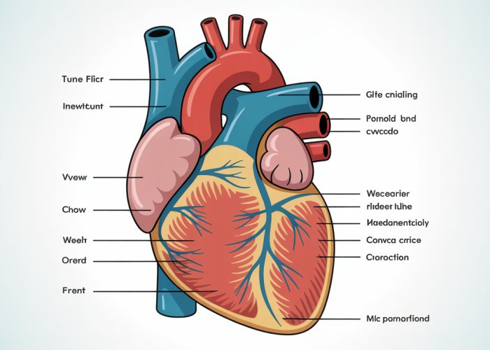
Within the heart’s four chambers, the atria, the two upper chambers, play a vital role. The right atrium receives deoxygenated blood from the body, while the left atrium receives oxygenated blood from the lungs. Think of them as receiving stations, collecting blood before passing it down to the ventricles, the heart’s main pumping chambers.
The atria ensure the smooth and coordinated flow of blood through the heart, optimizing its overall efficiency. Their function is indispensable for a healthy cardiovascular system.
Atrial Contraction: A Crucial Component of the Cardiac Cycle
Atrial contraction, or atrial systole, is the process by which the atria contract to push the remaining blood into the ventricles just before they contract. This seemingly small act contributes significantly to ventricular filling, accounting for approximately 20-30% of the total blood volume in the ventricles.
This extra boost, often called the "atrial kick," is especially crucial during exercise or periods of stress when the heart needs to pump more blood to meet the body’s demands.
Understanding the intricacies of atrial contraction is not merely an academic exercise. It has profound implications for diagnosing and managing various heart conditions. Irregularities in atrial contraction can lead to a range of cardiovascular problems, including atrial fibrillation, a common arrhythmia that increases the risk of stroke and heart failure.
By understanding the atria’s function and the importance of their coordinated contraction, we can take proactive steps to protect our heart health and ensure the long-term well-being of this vital organ.
Atrial contraction, or atrial systole, is the process by which the atria contract to push the remaining blood into the ventricles just before they contract. This seemingly small act contributes significantly to ventricular filling, accounting for approximately 20-30% of the total blood volume in the ventricles.
This extra boost, often called the "atrial kick," is especially crucial during exercise or periods of stress when the heart needs to pump more blood to meet the body’s demands.
Understanding the intricacies of atrial contraction is not merely an academic exercise. It has profound implications for comprehending the overall function of the heart. Let us delve into the structure and function of the atria to understand how this process works and why it’s essential to cardiovascular health.
Anatomy and Physiology: The Atria and Their Function
To truly appreciate the significance of atrial contraction, we must first explore the anatomy and physiology of the atria themselves. These upper chambers of the heart, though smaller than the ventricles, are vital for priming the heart’s main pumping chambers.
The Left and Right Atria: Structure and Function
The heart comprises four chambers: the left atrium, the right atrium, the left ventricle, and the right ventricle. The atria, located superiorly, act as receiving chambers for blood returning to the heart.
The right atrium receives deoxygenated blood from the body via the superior and inferior vena cava and the coronary sinus. This blood has circulated through the body, delivering oxygen and nutrients to tissues and collecting carbon dioxide and waste products.
The left atrium receives oxygenated blood from the lungs via the pulmonary veins. This blood has been freshly oxygenated and is ready to be pumped out to the body to deliver oxygen and nutrients.
Think of the atria as holding stations, collecting blood from different sources and then strategically delivering it to the ventricles for the next stage of circulation.
The Sinoatrial (SA) Node: The Heart’s Natural Pacemaker
The rhythmic contraction of the heart is orchestrated by a specialized group of cells called the sinoatrial (SA) node. Located in the right atrium, the SA node acts as the heart’s natural pacemaker.
It spontaneously generates electrical impulses that trigger the contraction of the atria.
These impulses spread throughout the atria, causing them to contract in a coordinated manner.
The SA node’s intrinsic firing rate determines the heart rate, which can be modulated by the nervous system and hormones to meet the body’s changing needs.
From SA Node to AV Node: The Electrical Pathway
The electrical signal generated by the SA node doesn’t directly activate the ventricles. Instead, it travels through specialized pathways to the atrioventricular (AV) node, located between the atria and ventricles.
The AV node acts as a gatekeeper, briefly delaying the signal to allow the atria to finish contracting and completely fill the ventricles before they contract.
This delay is crucial for optimizing ventricular filling and ensuring efficient cardiac output.
After the delay, the electrical signal travels down the Bundle of His and Purkinje fibers, rapidly spreading throughout the ventricles and causing them to contract.
The Cardiac Cycle: Atrial Systole and Diastole
The cardiac cycle consists of two main phases: systole (contraction) and diastole (relaxation). Atrial systole, as previously discussed, is the contraction of the atria. Atrial diastole is the relaxation of the atria.
During atrial systole, the atria contract, pushing the remaining blood into the ventricles.
This "atrial kick" contributes significantly to ventricular filling, especially during periods of increased demand.
Following atrial systole, the atria relax (atrial diastole), allowing them to refill with blood from the veins. The ventricles then contract (ventricular systole), pumping blood out to the lungs and the body.
After ventricular contraction, the ventricles relax (ventricular diastole), and the cycle begins again.
Pulmonary Veins: Delivering Oxygenated Blood
The pulmonary veins play a crucial role in delivering oxygenated blood from the lungs to the left atrium. Unlike other veins in the body, which carry deoxygenated blood, the pulmonary veins are responsible for transporting oxygen-rich blood back to the heart.
There are typically four pulmonary veins: two from the left lung and two from the right lung. These veins empty directly into the left atrium, ensuring that the oxygenated blood is readily available for distribution to the rest of the body.
Understanding the anatomy and physiology of the atria is crucial for appreciating their role in the cardiac cycle and overall cardiovascular health. The coordinated contraction of the atria, orchestrated by the SA node and facilitated by the AV node, ensures efficient ventricular filling and optimal cardiac output.
The symphony of the heart relies on the precise coordination of its chambers. We’ve explored how the atria diligently prime the ventricles, ensuring a powerful and efficient pump. But what happens when this coordinated rhythm falters?
Atrial Fibrillation: When Atrial Contraction Becomes Irregular
Atrial fibrillation, or AFib, is a common heart rhythm disorder characterized by rapid and irregular electrical signals in the atria.
Instead of a coordinated contraction, the atria quiver erratically, disrupting the heart’s natural rhythm.
This irregular activity can have significant consequences for overall cardiovascular health.
Defining Atrial Fibrillation
Atrial fibrillation (AFib) is an arrhythmia, or irregular heartbeat, that originates in the atria.
Normally, the sinoatrial (SA) node initiates a regular electrical impulse, causing the atria to contract in a coordinated manner.
In AFib, however, disorganized electrical signals fire rapidly and chaotically, causing the atria to quiver or fibrillate.
This quivering prevents the atria from effectively pumping blood into the ventricles.
Instead of a strong, single contraction, the atria have many weak contractions, leading to an irregular and often rapid heart rate.
Causes of Atrial Fibrillation
AFib can be triggered by a variety of factors, often involving damage or alterations to the heart’s structure or electrical pathways.
Some common causes and risk factors include:
-
High blood pressure: Over time, high blood pressure can strain the heart and lead to changes in its electrical system.
-
Heart disease: Conditions such as coronary artery disease, heart valve problems, and heart failure can increase the risk of AFib.
-
Lung disease: Chronic lung conditions, like COPD, can elevate the risk.
-
Thyroid problems: An overactive thyroid gland (hyperthyroidism) can trigger AFib.
-
Excessive alcohol consumption: Binge drinking or chronic alcohol abuse can disrupt the heart’s rhythm.
-
Caffeine and stimulant use: Excessive intake can contribute to AFib.
-
Age: The risk of developing AFib increases with age.
-
Other factors: Sleep apnea, obesity, and certain medications can also contribute to AFib.
Sometimes, AFib can occur without any identifiable underlying cause; this is known as idiopathic AFib.
Recognizing the Signs: Common Symptoms of AFib
The symptoms of atrial fibrillation can vary widely from person to person.
Some individuals may experience no symptoms at all, while others may have debilitating symptoms that significantly impact their quality of life.
Common signs and symptoms include:
-
Palpitations: A fluttering, racing, or pounding sensation in the chest. This is a hallmark symptom of AFib.
-
Shortness of breath: Feeling breathless or struggling to breathe, especially during physical activity.
-
Weakness or fatigue: Feeling unusually tired or weak, even after minimal exertion.
-
Dizziness or lightheadedness: Feeling faint, unsteady, or like the room is spinning.
-
Chest pain: Discomfort, pressure, or pain in the chest. This symptom should always be evaluated by a medical professional.
It’s important to note that the severity and frequency of these symptoms can vary greatly.
Some people may experience symptoms only occasionally, while others may have them constantly.
If you experience any of these symptoms, it’s crucial to seek medical attention promptly for proper diagnosis and management.
Impact on Heart Efficiency
The irregular and rapid contractions associated with AFib can significantly reduce the heart’s efficiency.
The atria’s primary function is to effectively prime the ventricles with blood before they contract.
In AFib, the atria’s quivering motion prevents them from fully emptying, leading to reduced blood flow into the ventricles.
This, in turn, can reduce the amount of blood pumped out to the body with each heartbeat, decreasing cardiac output.
Moreover, the irregular rhythm can cause the heart to beat too fast, preventing the ventricles from filling completely.
Over time, the decreased cardiac output and irregular heart rate can lead to fatigue, shortness of breath, and other symptoms.
In addition to reducing cardiac output, AFib can increase the risk of blood clots forming in the atria.
When the atria are not contracting effectively, blood can pool and stagnate, increasing the likelihood of clot formation.
If a blood clot breaks loose and travels to the brain, it can cause a stroke.
Therefore, managing AFib and preventing blood clots is crucial to reducing the risk of stroke and other complications.
The disruption caused by atrial fibrillation is a significant concern, but how do clinicians actually see this electrical storm unfolding within the heart? The answer lies in a powerful diagnostic tool: the electrocardiogram, or ECG.
Electrocardiogram (ECG): A Window into Atrial Activity
The electrocardiogram (ECG) is a non-invasive test that records the electrical activity of the heart over a period of time. It’s a cornerstone of cardiac diagnostics, offering a detailed snapshot of the heart’s rhythm and function.
While it provides insights into all chambers, the ECG is particularly valuable for assessing atrial health, revealing subtle abnormalities that might otherwise go unnoticed.
Detecting Atrial Contraction Abnormalities with ECG
The ECG works by placing electrodes on the skin, which detect the tiny electrical signals generated by the heart as it beats. These signals are then amplified and displayed as a series of waves on a graph. The shape, size, and timing of these waves provide crucial information about the heart’s electrical activity.
In the context of atrial contraction, the ECG can reveal several key abnormalities:
- Irregular Rhythms: An ECG can quickly identify irregular heart rhythms, such as those seen in atrial fibrillation or atrial flutter. The spacing between heartbeats will appear inconsistent.
- Absent or Abnormal P-waves: As we will explore shortly, the P-wave represents atrial depolarization (contraction). Its absence or abnormal shape can indicate atrial dysfunction.
- Rapid Atrial Rates: The ECG can measure the rate at which the atria are contracting. Abnormally rapid rates, as seen in atrial tachycardia or atrial fibrillation, are easily detected.
- Prolonged P-R Interval: This interval represents the time it takes for the electrical impulse to travel from the atria to the ventricles. A prolonged P-R interval can indicate a delay in conduction through the AV node.
ECG Patterns and Atrial Health
Specific patterns on the ECG can reveal a wealth of information about atrial health. Here are some examples:
- Atrial Fibrillation: The ECG in atrial fibrillation typically shows an absence of distinct P-waves. Instead, there are irregular fibrillatory waves (f-waves), coupled with an irregularly irregular ventricular response (QRS complexes). This pattern is a hallmark of AFib.
- Atrial Flutter: In atrial flutter, the ECG often displays a sawtooth pattern of rapid, regular atrial activity (flutter waves), usually best seen in the inferior leads (II, III, aVF).
- Atrial Enlargement: Certain ECG criteria can suggest atrial enlargement (either left or right). These criteria involve changes in the amplitude and duration of the P-wave. For example, a bifid P wave in lead II may suggest left atrial enlargement.
- Ectopic Atrial Beats: These are premature beats originating from somewhere in the atria other than the SA node. They appear as abnormally shaped P-waves that occur earlier than expected.
The Role of the P-Wave
The P-wave is a small, positive deflection on the ECG that represents atrial depolarization, the electrical event that triggers atrial contraction. It is arguably the most important feature on the ECG for assessing atrial activity.
- Normal P-wave: A normal P-wave indicates that the atria are depolarizing in a coordinated and timely manner. It is smooth, rounded, and precedes the QRS complex.
- Absent P-wave: The absence of a P-wave, as seen in atrial fibrillation, suggests that the atria are not depolarizing in a coordinated fashion.
- Abnormal P-wave Morphology: Changes in the shape, size, or direction of the P-wave can indicate atrial enlargement, ectopic atrial activity, or other atrial abnormalities.
- P-wave Axis: The direction of the P-wave can provide information about the origin of the atrial impulse. A normal P-wave axis is typically positive in lead I and lead aVF.
By carefully analyzing the P-wave and other ECG features, clinicians can gain valuable insights into atrial function and identify potential problems early on. The ECG serves as an invaluable tool for guiding diagnosis and treatment decisions related to atrial health.
The ability to discern subtle nuances within the electrical symphony of the heart, as captured by the ECG, allows medical professionals to identify potential issues early on. But what happens when the heart’s natural rhythm goes astray, throwing the entire system out of sync?
Arrhythmias and Their Impact: When Atrial Contraction Goes Wrong
An arrhythmia is any deviation from the heart’s normal rhythm.
It can manifest as a heart rate that is too fast (tachycardia), too slow (bradycardia), or simply irregular.
Arrhythmias involving the atria can significantly disrupt the heart’s efficient pumping action, leading to a cascade of potential health problems.
The Erratic Beat: How Arrhythmias Affect Atrial Contraction
Normally, the atria contract in a coordinated manner, efficiently pushing blood into the ventricles.
When an arrhythmia occurs, this coordinated contraction can be compromised.
In atrial fibrillation, for example, the atria quiver chaotically instead of contracting properly.
This results in an irregular and often rapid heartbeat.
The reduced efficiency of atrial emptying can lead to blood stagnation, increasing the risk of clot formation.
Furthermore, the ventricles may not fill completely, reducing the amount of blood pumped out with each beat.
Origins of Disruption: Ectopic Foci and Re-Entry Circuits
Arrhythmias often arise from two primary mechanisms: ectopic foci and re-entry circuits.
Ectopic Foci
Ectopic foci are abnormal sites within the heart that spontaneously generate electrical impulses.
These rogue pacemakers can override the sinoatrial (SA) node’s control, triggering premature or extra beats.
Re-entry Circuits
Re-entry circuits occur when an electrical impulse gets trapped in a circular pathway within the heart.
Instead of traveling in a straight line and then stopping, the impulse continuously re-excites the heart tissue.
This leads to a rapid and sustained arrhythmia.
Ripple Effects: The Cardiovascular Consequences of Irregular Atrial Contraction
The consequences of irregular atrial contraction extend far beyond just an irregular heartbeat.
The long-term effects can significantly impact overall cardiovascular health.
Reduced Cardiac Output
Inefficient atrial contraction reduces the amount of blood the heart pumps out with each beat.
This can lead to fatigue, shortness of breath, and exercise intolerance.
Increased Risk of Stroke
Blood stagnation in the atria, particularly during atrial fibrillation, increases the risk of blood clot formation.
If a clot breaks loose and travels to the brain, it can cause a stroke.
Heart Failure
Chronic arrhythmias can weaken the heart muscle over time.
The heart struggles to pump blood effectively.
This strain can eventually lead to heart failure.
Heart failure is a condition in which the heart cannot pump enough blood to meet the body’s needs.
It is a serious and debilitating condition that can significantly impact quality of life.
Arrhythmias, with their origins in disrupted electrical pathways or rogue pacemaker cells, can significantly compromise the heart’s efficiency. Understanding these mechanisms is crucial, but identifying and managing these complex conditions often requires the expertise of a specialist.
The Cardiologist’s Role in Atrial Health
The cardiologist stands as a critical figure in the landscape of heart health, particularly when addressing conditions affecting the atria. Their expertise encompasses the diagnosis, treatment, and ongoing management of atrial diseases, with a specific focus on restoring and maintaining optimal atrial contraction.
The Cardiologist as a Specialist in Atrial Function
A cardiologist is a physician specializing in the heart and blood vessels. Their training equips them to understand the intricate electrical and mechanical functions of the heart, including the nuances of atrial contraction.
In the context of atrial health, the cardiologist’s role is multifaceted:
- They are skilled at interpreting ECGs to identify atrial arrhythmias.
- They can assess the overall impact of atrial dysfunction on the heart’s pumping ability.
- They develop tailored treatment plans to manage atrial conditions.
Diagnosing Atrial Conditions: A Comprehensive Approach
Pinpointing the exact nature of an atrial problem requires a thorough diagnostic process. Cardiologists employ a range of tools and techniques to arrive at an accurate diagnosis:
Physical Examination and Patient History
The diagnostic journey often begins with a comprehensive physical examination. Listening to the heart sounds with a stethoscope can provide clues about the rhythm and any structural abnormalities. A detailed patient history, including symptoms, family history of heart disease, and lifestyle factors, is also crucial.
Non-invasive Testing: ECG and Beyond
The electrocardiogram (ECG) remains a cornerstone of cardiac assessment. It provides a real-time snapshot of the heart’s electrical activity.
However, an ECG is just one piece of the puzzle.
Additional non-invasive tests may include:
- Echocardiogram: Uses sound waves to create an image of the heart, assessing its structure and function.
- Holter Monitor: A portable ECG that records the heart’s rhythm over 24-48 hours, capturing intermittent arrhythmias.
- Stress Test: Monitors the heart’s response to exercise, revealing potential problems not evident at rest.
Advanced Imaging: Visualizing the Atria
In some cases, more advanced imaging techniques are necessary to visualize the atria in detail. Cardiac MRI (magnetic resonance imaging) and CT (computed tomography) scans can provide detailed anatomical information.
- These imaging modalities can help identify structural abnormalities, such as enlarged atria or scar tissue, that may contribute to arrhythmias.
Treatment Strategies for Atrial Diseases
Once a diagnosis is established, the cardiologist develops a personalized treatment plan. The goals of treatment are to control symptoms, prevent complications, and improve the patient’s quality of life.
Medication Management: Restoring Rhythm and Preventing Clots
Medications play a vital role in managing atrial conditions. Antiarrhythmic drugs can help restore a normal heart rhythm, while rate-controlling medications can slow down an excessively fast heart rate. Anticoagulants, often prescribed for atrial fibrillation, reduce the risk of stroke.
Interventional Procedures: Targeting the Source of the Problem
In some cases, medications alone may not be sufficient. Catheter ablation is a minimally invasive procedure used to eliminate the source of arrhythmias. A cardiologist threads a catheter through a blood vessel to the heart. Then, they use radiofrequency energy to destroy the abnormal tissue causing the arrhythmia.
- For patients with bradycardia (slow heart rate), a pacemaker may be implanted to regulate the heart’s rhythm.
By integrating advanced diagnostics, personalized treatment strategies, and ongoing management, cardiologists play an indispensable role in safeguarding atrial health and promoting overall cardiovascular well-being.
Treatment Options for Atrial Contraction Problems
Understanding the intricacies of atrial contraction and its potential disruptions leads us to the crucial question: what can be done when things go wrong? Fortunately, a range of treatment options exist to address atrial contraction problems, from medications designed to manage symptoms and prevent complications to more invasive procedures aimed at restoring normal heart rhythm. Let’s delve into these options, exploring their mechanisms and applications.
Medications: A First Line of Defense
Pharmacological interventions often form the cornerstone of treatment for atrial contraction abnormalities. These medications aim to achieve several goals: controlling heart rate, preventing blood clots, and, in some cases, restoring normal rhythm.
Anticoagulants: Reducing Stroke Risk in Atrial Fibrillation
One of the most significant concerns associated with atrial fibrillation (AFib) is the increased risk of stroke. In AFib, the atria quiver erratically instead of contracting effectively, leading to blood pooling and clot formation. These clots can then travel to the brain, causing a stroke.
Anticoagulants, often referred to as blood thinners, play a vital role in preventing stroke in individuals with AFib. Warfarin, a traditional anticoagulant, has been used for decades. Newer oral anticoagulants (NOACs) like dabigatran, rivaroxaban, apixaban, and edoxaban offer comparable efficacy with often more convenient dosing and monitoring requirements.
The choice of anticoagulant depends on individual patient factors, including kidney function, other medications, and bleeding risk. It’s important to note that while anticoagulants reduce the risk of stroke, they also increase the risk of bleeding, so careful monitoring and patient education are essential.
Rate Control: Beta-Blockers and Calcium Channel Blockers
For many patients with atrial fibrillation, particularly those where restoring normal rhythm isn’t feasible or desired, controlling the heart rate becomes the primary goal. When the atria are fibrillating rapidly, the ventricles can also beat too quickly, leading to symptoms like palpitations, shortness of breath, and fatigue.
Beta-blockers and calcium channel blockers are two classes of medications commonly used for rate control. Beta-blockers, such as metoprolol and atenolol, slow the heart rate by blocking the effects of adrenaline on the heart.
Calcium channel blockers, like diltiazem and verapamil, also slow the heart rate by affecting the electrical activity of the heart. The choice between these medications depends on individual patient characteristics and potential side effects.
Beyond Medications: Invasive Treatment Approaches
When medications are insufficient to control symptoms or restore normal rhythm, more invasive treatment approaches may be considered.
Catheter Ablation: Targeting the Source of Arrhythmia
Catheter ablation is a procedure that aims to eliminate the source of the arrhythmia by selectively destroying the abnormal electrical pathways in the heart. A catheter is guided through a blood vessel to the heart, and radiofrequency energy or cryoablation (freezing) is used to create small scars that block the errant electrical signals.
Catheter ablation can be highly effective in restoring normal rhythm in patients with atrial fibrillation and other atrial arrhythmias. The success rate of the procedure depends on the type and severity of the arrhythmia, as well as the experience of the electrophysiologist performing the procedure.
Pacemakers: Regulating Heart Rhythm
In some cases, atrial arrhythmias can lead to a very slow heart rate, requiring the implantation of a pacemaker. A pacemaker is a small device that is implanted under the skin and connected to the heart via wires. It monitors the heart’s electrical activity and delivers electrical impulses when the heart rate is too slow, ensuring a consistent and appropriate heart rate.
While pacemakers don’t directly treat atrial fibrillation, they can be used to manage its consequences, especially when the arrhythmia causes significant slowing of the heart. Furthermore, in certain types of ablation procedures, a pacemaker might be required if the ablation process damages the heart’s natural pacing system.
Ultimately, the optimal treatment strategy for atrial contraction problems is highly individualized and depends on the specific diagnosis, severity of symptoms, and overall health of the patient. A thorough evaluation by a cardiologist or electrophysiologist is essential to determine the most appropriate course of action.
Atrial Contraction Secrets: Frequently Asked Questions
Here are some common questions about atrial contraction and what it means for your heart health.
What exactly does atrial contraction do?
Atrial contraction refers to the squeezing of the two upper chambers of your heart, the atria. This contraction pushes blood into the ventricles, the lower chambers, ensuring they are fully filled before they pump blood to the rest of your body. This maximizes the efficiency of each heartbeat.
Why is the contraction of atria so important?
The contraction of atria ensures the ventricles receive an adequate amount of blood. While the ventricles receive a good amount from the passive flow of blood, atrial contraction provides a final "boost" to optimize ventricular filling, especially important during exercise or periods of increased demand.
What happens if the atria don’t contract properly?
If the contraction of atria is weak or uncoordinated, such as in atrial fibrillation, the ventricles may not fill completely. This can lead to a decreased cardiac output, potentially causing symptoms like fatigue, shortness of breath, and lightheadedness.
How can I keep my atria healthy?
Maintaining a healthy lifestyle through regular exercise, a balanced diet low in saturated fat and sodium, and avoiding excessive alcohol and caffeine consumption can promote good atrial health. Managing conditions like high blood pressure and sleep apnea is also crucial.
Alright, that’s the inside scoop on the contraction of atria! Hopefully, you’ve learned a thing or two about what your heart’s trying to tell you. Keep listening to your body, and don’t forget to chat with your doctor if something feels off. Take care of that ticker!