Understanding the aqueous humor is essential for comprehending intraocular pressure, a critical factor monitored during routine glaucoma screenings. The precise aqueous humor location, found within both the anterior and posterior chambers of the eye, directly influences this pressure. Its production by the ciliary body and subsequent flow through the trabecular meshwork are directly linked to maintaining healthy vision, further reinforcing the importance of the aqueous humor location in overall eye health.
The eye, a marvel of biological engineering, relies on a delicate interplay of structures and fluids to capture the world around us. Among these essential components is the aqueous humor, a clear, watery fluid that resides within the anterior segment of the eye. Often overlooked, this fluid is crucial for maintaining ocular health and enabling clear vision.
Defining Aqueous Humor and Its Significance
Aqueous humor is not simply "eye water". It is a complex solution containing nutrients, electrolytes, and other essential components.
Its primary function is to nourish the avascular (blood vessel-free) structures of the eye, namely the cornea and the lens, delivering vital substances that these tissues cannot obtain directly from the bloodstream.
Beyond nutrition, aqueous humor plays a critical role in maintaining intraocular pressure (IOP), the pressure inside the eye. This pressure is essential for maintaining the globe’s shape and ensuring proper optical function. Without adequate IOP, the eye would collapse, and vision would be severely compromised.
Purpose: Location and Role of Aqueous Humor Explored
This article aims to shed light on the aqueous humor, exploring its specific location within the eye and its multifaceted roles in maintaining ocular health. By understanding where this fluid resides and what functions it performs, we can better appreciate its significance. We aim to provide a comprehensive understanding accessible to all readers, regardless of their background in medicine or ophthalmology.
Potential Problems: Aqueous Humor Imbalance and Glaucoma
The delicate balance of aqueous humor production and drainage is crucial for maintaining healthy IOP. When this balance is disrupted, serious eye conditions can arise. Perhaps the most well-known of these is glaucoma, a leading cause of irreversible blindness worldwide.
Glaucoma often results from impaired drainage of aqueous humor, leading to a buildup of pressure inside the eye. This elevated pressure can damage the optic nerve, gradually leading to vision loss.
While glaucoma is perhaps the most significant condition linked to aqueous humor imbalance, other ocular diseases can also be affected by disruptions in its production, flow, or composition. Understanding the potential problems associated with aqueous humor is crucial for preventative eye care and early intervention.
The aqueous humor’s role cannot be fully appreciated without first understanding the intricate landscape of the eye itself. Its production, circulation, and eventual drainage are all intimately linked to the specific structures within this complex organ.
Anatomy of the Eye: A Roadmap for Aqueous Humor
The eye, while seemingly simple in its outward appearance, is a sophisticated system of interconnected components. Each part contributes to the overall function of vision, and several play critical roles in the dynamics of aqueous humor.
Key Eye Structures: A Brief Overview
-
Cornea: The clear, dome-shaped front surface of the eye. It acts as the primary refractive surface, bending light as it enters the eye. The cornea receives nutrients from the aqueous humor.
-
Lens: A transparent, biconvex structure located behind the iris. It focuses light onto the retina, allowing for clear vision at varying distances. Like the cornea, the lens is avascular and relies on aqueous humor for nourishment.
-
Iris: The colored part of the eye, which controls the amount of light entering the eye by adjusting the size of the pupil.
-
Ciliary Body: Located behind the iris, this structure produces aqueous humor and contains the ciliary muscle, which controls the shape of the lens for accommodation (focusing).
-
Optic Nerve: Transmits visual information from the retina to the brain.
These structures, among others, work in concert to enable sight.
However, for our discussion on aqueous humor, the spotlight shines on the anterior segment, particularly the anterior and posterior chambers.
Anterior and Posterior Chambers: The Aqueous Humor’s Domain
The anterior segment of the eye is the area in front of the lens. Within this segment lie two crucial spaces: the anterior and posterior chambers.
Defining the Boundaries
The posterior chamber is a small space located behind the iris and in front of the lens. It extends from the back of the iris to the front of the lens and is also bordered by the ciliary body.
This is the primary site of aqueous humor production.
The anterior chamber is the space between the cornea and the iris. It is larger than the posterior chamber and is filled with aqueous humor that has flowed from the posterior chamber.
The Relationship Between the Chambers
These two chambers are directly connected and form a continuous pathway for the aqueous humor.
The aqueous humor, produced by the ciliary body in the posterior chamber, flows through the pupil (the opening in the iris) into the anterior chamber.
From there, it circulates and eventually drains out of the eye through the trabecular meshwork (more on this later).
Why Eye Anatomy Matters: A Foundation for Understanding
Understanding the anatomical relationships within the eye is paramount to grasping the dynamics of aqueous humor. Knowing the location of the ciliary body, the boundaries of the anterior and posterior chambers, and the flow path through the pupil is essential for comprehending how this fluid nourishes the eye and maintains proper intraocular pressure. Without this foundational knowledge, discussions about glaucoma and other aqueous humor-related conditions become significantly more difficult to follow.
Anterior and posterior chambers are the main areas that we have been shining light on. However, where exactly does the aqueous humor reside, and how does it navigate this intricate ocular landscape? Understanding its precise location and flow path is crucial to appreciating its role in maintaining eye health.
Location, Location, Location: Pinpointing Aqueous Humor’s Residence
The journey of aqueous humor within the eye is a carefully orchestrated flow, starting with its production and ending with its drainage. Let’s trace its path step by step.
The Ciliary Body: The Source of Aqueous Humor
The ciliary body, located behind the iris, is the powerhouse responsible for producing aqueous humor.
Specialized cells within the ciliary body, specifically the non-pigmented ciliary epithelium, actively secrete the fluid into the posterior chamber.
This process is not simply a passive leakage; it’s an active transport mechanism that carefully controls the composition of the aqueous humor.
From Posterior to Anterior: Navigating the Pupil
Once produced in the posterior chamber, the aqueous humor embarks on its journey forward.
It flows through the pupil, the opening in the center of the iris, into the anterior chamber.
This transition from the posterior to anterior chamber is a critical step in the fluid’s circulation.
The Anterior and Posterior Chambers: Aqueous Humor’s Primary Residence
The anterior and posterior chambers are the primary locations where aqueous humor resides and performs its essential functions.
These two chambers, while distinct, are intimately connected by the pupil, allowing for continuous fluid circulation.
The constant flow ensures that nutrients are delivered and waste products are removed efficiently.
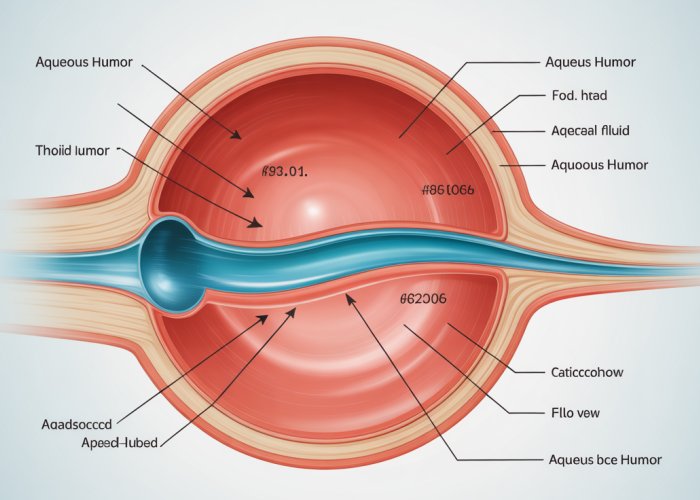
Visualizing the Flow Path
To fully grasp the journey of aqueous humor, a visual aid can be invaluable.
A diagram illustrating the eye’s anatomy, with arrows indicating the direction of aqueous humor flow, can provide a clearer understanding of its circulation.
Such a visual representation would highlight the production in the ciliary body, the passage through the pupil, and the presence in both anterior and posterior chambers, leading ultimately to the drainage pathways.
Anterior and posterior chambers are the main areas that we have been shining light on. However, where exactly does the aqueous humor reside, and how does it navigate this intricate ocular landscape? Understanding its precise location and flow path is crucial to appreciating its role in maintaining eye health.
The Crucial Role: Functions of Aqueous Humor
Beyond its location, the aqueous humor’s true significance lies in the multifaceted roles it performs to maintain the health and functionality of the eye. This clear fluid is far more than just a space filler; it is a dynamic medium responsible for nutrient delivery, waste removal, and the maintenance of optimal intraocular pressure (IOP). These functions are critical to preserving vision and overall eye well-being.
Nourishment of Avascular Tissues
One of the primary responsibilities of the aqueous humor is to provide essential nutrients to the avascular structures of the eye, namely the lens and the cornea. Unlike many other tissues in the body, these structures lack a direct blood supply.
Therefore, they rely entirely on the aqueous humor to deliver glucose, amino acids, and other vital nutrients required for their metabolic processes and overall health. Without this continuous supply, the lens and cornea would suffer from malnutrition, leading to impaired function and potential vision loss.
Waste Removal and Detoxification
In addition to delivering nutrients, the aqueous humor also acts as a crucial waste removal system for the eye. As the lens and cornea metabolize nutrients, they produce waste products that must be efficiently eliminated.
The aqueous humor collects these metabolic byproducts and carries them away, preventing their build-up within the eye. This detoxification process is essential for maintaining a clear optical pathway and preventing cellular damage.
Maintaining Intraocular Pressure (IOP)
Perhaps the most well-known function of the aqueous humor is its role in maintaining intraocular pressure (IOP). IOP refers to the fluid pressure inside the eye, which is critical for maintaining the eye’s shape and overall structural integrity.
The continuous production and drainage of aqueous humor create a delicate balance that keeps the IOP within a normal range. This pressure supports the eyeball, preventing it from collapsing or distorting, which could lead to blurred vision or other complications.
IOP and Ocular Health: A Delicate Balance
The IOP maintained by the aqueous humor is not just about physical shape; it is also intimately linked to the health of the optic nerve, which transmits visual information from the eye to the brain. When the IOP is too high, it can damage the delicate nerve fibers of the optic nerve, leading to a condition known as glaucoma.
Glaucoma is a leading cause of irreversible blindness worldwide, and elevated IOP is a major risk factor. Conversely, low IOP (hypotony) can also cause issues, such as choroidal effusions or retinal detachment.
Therefore, maintaining the right balance of aqueous humor production and drainage is critical in preserving sight and preventing potentially blinding diseases.
The continuous production of aqueous humor necessitates a carefully orchestrated drainage system to maintain a stable intraocular pressure. The primary route for this outflow involves two critical structures: the trabecular meshwork and Schlemm’s canal, working in conjunction with the anterior chamber angle. Disruptions to this intricate drainage pathway can have significant consequences for eye health, most notably in the development of glaucoma.
The Drainage System: Trabecular Meshwork and Schlemm’s Canal
Understanding how aqueous humor exits the eye is just as vital as understanding how it’s produced. The trabecular meshwork, Schlemm’s canal, and the anterior chamber angle form an intricate outflow system. When this system is compromised, it can lead to serious ocular conditions.
The Trabecular Meshwork: A Filter System
The trabecular meshwork is a complex, sponge-like tissue located in the anterior chamber angle. It acts as the primary filter for aqueous humor as it exits the eye.
This meshwork consists of a series of interconnected channels and spaces, providing resistance to the flow of fluid. This resistance is essential for maintaining a healthy intraocular pressure (IOP).
The structure of the trabecular meshwork is quite intricate, consisting of several layers with varying pore sizes. This complex architecture allows it to effectively filter out debris and particles from the aqueous humor, preventing them from entering Schlemm’s canal.
Schlemm’s Canal: The Collection Channel
After passing through the trabecular meshwork, the aqueous humor enters Schlemm’s canal, a circular channel that encircles the cornea. Schlemm’s canal acts as a collection vessel.
From here, the aqueous humor is directed into a network of collector channels and eventually into the episcleral veins, rejoining the systemic circulation. The endothelium lining Schlemm’s canal plays a vital role in regulating outflow.
Dysfunction of this endothelium can significantly impact the drainage process. The canal’s integrity is crucial for efficient fluid removal.
The Anterior Chamber Angle: Where It All Converges
The anterior chamber angle is the anatomical space where the cornea and iris meet. This angle is critically important because it houses both the trabecular meshwork and the entrance to Schlemm’s canal.
Defining the Angle
The anterior chamber angle isn’t just a passive space; it’s a dynamic area where the aqueous humor gains access to the primary drainage pathway.
Its openness and structural integrity directly impact the efficiency of aqueous outflow. A narrow or closed angle can physically obstruct the trabecular meshwork, hindering drainage.
Angle’s Affect on Drainage
The angle’s configuration significantly affects aqueous humor drainage. An open, wide angle allows for unimpeded flow into the trabecular meshwork, while a narrow or closed angle restricts this flow.
Conditions that cause the iris to press against the cornea, such as angle-closure glaucoma, can physically block the trabecular meshwork, leading to a rapid increase in IOP. Gonioscopy, a diagnostic procedure, is used to examine the angle’s structure.
Impact of Drainage Issues on Intraocular Pressure
When the outflow of aqueous humor is obstructed, the fluid accumulates within the eye. This leads to an elevation of intraocular pressure (IOP).
This increased pressure can damage the optic nerve, leading to glaucoma, a leading cause of irreversible blindness worldwide.
The relationship between drainage issues and increased IOP is direct and significant. Any factor that impedes the flow of aqueous humor through the trabecular meshwork or Schlemm’s canal will inevitably lead to a rise in IOP. Early detection and management of drainage issues are crucial in preventing glaucomatous damage.
After passing through the trabecular meshwork and entering Schlemm’s canal, the aqueous humor embarks on its final journey, ultimately rejoining the systemic circulation. However, what happens when this carefully regulated outflow process encounters an obstruction? The consequences can be significant, leading to a buildup of pressure within the eye and potentially devastating effects on vision.
Aqueous Humor Imbalance: Understanding Glaucoma and Other Conditions
A delicate balance governs the production and drainage of aqueous humor. When this equilibrium is disrupted, the consequences can be detrimental to eye health. The most notable condition arising from such an imbalance is glaucoma, a leading cause of irreversible blindness worldwide.
Glaucoma: The Silent Thief of Sight
Glaucoma isn’t a single disease, but rather a group of optic neuropathies characterized by progressive damage to the optic nerve, often associated with elevated intraocular pressure (IOP). This pressure increase typically stems from impaired aqueous humor drainage, causing a gradual loss of peripheral vision that can eventually lead to complete blindness if left untreated.
The insidious nature of glaucoma lies in its often asymptomatic early stages. Individuals may not notice any changes in their vision until significant damage has already occurred. This is why regular eye exams are crucial for early detection and intervention.
Types of Glaucoma Related to Aqueous Humor Dynamics
Several types of glaucoma are directly linked to issues with aqueous humor flow:
Open-Angle Glaucoma
Open-angle glaucoma is the most common form. In this condition, the anterior chamber angle remains open, but the trabecular meshwork gradually becomes less efficient at draining aqueous humor. The exact cause of this reduced outflow is not fully understood, but factors like age, genetics, and other underlying medical conditions may play a role.
Angle-Closure Glaucoma
Angle-closure glaucoma, also known as closed-angle or narrow-angle glaucoma, occurs when the iris physically blocks the drainage angle, preventing aqueous humor from reaching the trabecular meshwork. This can happen suddenly (acute angle-closure) or gradually over time (chronic angle-closure). Acute angle-closure is a medical emergency requiring immediate treatment to prevent rapid vision loss.
Normal-Tension Glaucoma
Normal-tension glaucoma is a unique form where optic nerve damage occurs despite having normal IOP. The exact mechanism behind this is still under investigation, but it’s believed that the optic nerve may be more susceptible to damage in some individuals, even at normal pressure levels. Vascular factors and other systemic conditions may also contribute.
Beyond Glaucoma: Other Conditions Affected by Aqueous Humor
While glaucoma is the most well-known condition associated with aqueous humor imbalance, other ocular problems can also arise from disruptions in its production or drainage.
-
Uveitis: Inflammation of the uveal tract (iris, ciliary body, and choroid) can affect aqueous humor production and composition. Inflammatory cells and proteins can accumulate in the aqueous humor, leading to increased IOP and other complications.
-
Hyphema: Bleeding into the anterior chamber, often caused by trauma, can obstruct aqueous humor outflow and raise IOP.
-
Iritis: Inflammation of the iris can lead to the formation of adhesions that obstruct the flow of aqueous humor through the pupil, potentially leading to increased IOP.
-
Secondary Glaucomas: Various conditions, such as steroid use, eye injuries, or certain medical conditions, can lead to secondary glaucomas by affecting aqueous humor dynamics.
After passing through the trabecular meshwork and entering Schlemm’s canal, the aqueous humor embarks on its final journey, ultimately rejoining the systemic circulation. However, what happens when this carefully regulated outflow process encounters an obstruction? The consequences can be significant, leading to a buildup of pressure within the eye and potentially devastating effects on vision.
Diagnosis and Treatment: Proactive Steps for Preserving Vision
The intricacies of aqueous humor dynamics underscore the need for diligent eye care. Because many conditions related to aqueous humor imbalance, particularly glaucoma, present with minimal or no symptoms in their early stages, proactive measures are paramount. Regular eye examinations are not merely recommended; they are essential for safeguarding your sight.
The Vital Role of Regular Eye Exams
Think of regular eye exams as preventative maintenance for your vision. These check-ups, conducted by an ophthalmologist (a medical doctor specializing in eye care) or an optometrist (a healthcare professional trained to examine the eyes for visual defects and prescribe corrective lenses), play a crucial role in early detection.
Early detection allows for timely intervention, potentially preventing irreversible damage and vision loss.
These examinations encompass a comprehensive assessment of your eye health. This includes evaluating visual acuity, examining the structures of the eye, and measuring intraocular pressure (IOP).
Diagnostic Tools for Assessing Aqueous Humor Dynamics
Several sophisticated diagnostic tests are employed to evaluate aqueous humor drainage and IOP. These tests help eye care professionals identify potential problems and monitor the effectiveness of treatment strategies.
Tonometry: Measuring Intraocular Pressure
Tonometry is a fundamental test that measures the pressure inside your eye. Different methods exist, including:
- Goldmann Applanation Tonometry: Considered the gold standard, this method involves gently flattening a small area of the cornea to measure IOP.
- Non-Contact Tonometry (Air-Puff Tonometry): This method uses a puff of air to flatten the cornea, making it a quick and less invasive option.
Elevated IOP is a significant risk factor for glaucoma, but it’s important to remember that normal IOP doesn’t always rule out the condition. Some individuals may develop glaucoma with normal pressure, known as normal-tension glaucoma.
Gonioscopy: Examining the Drainage Angle
Gonioscopy allows the eye doctor to visualize the anterior chamber angle, where the iris and cornea meet. This angle is where aqueous humor drains out of the eye.
Using a special lens, the doctor can assess whether the angle is open or closed, and identify any abnormalities that might be obstructing the flow of aqueous humor. This is critical in diagnosing angle-closure glaucoma.
Visual Field Testing: Assessing Peripheral Vision
Visual field testing maps out the extent of your peripheral vision. This test can detect blind spots or areas of vision loss, which are characteristic of glaucoma.
Progressive vision loss, often starting in the periphery, is a hallmark of glaucoma, making visual field testing an essential tool for monitoring the disease’s progression.
Optical Coherence Tomography (OCT): Imaging the Optic Nerve
OCT is a non-invasive imaging technique that provides detailed cross-sectional images of the optic nerve and retina. It can detect subtle structural changes in the optic nerve, which may indicate early glaucoma damage, often before visual field loss is apparent. This early detection is key.
Treatment Options for Aqueous Humor-Related Conditions
Fortunately, various treatment options are available to manage conditions related to aqueous humor imbalance, particularly glaucoma. The primary goal of treatment is to lower IOP and prevent further damage to the optic nerve.
Medication: Eye Drops to Lower IOP
Eye drops are the most common initial treatment for glaucoma. These medications work by either decreasing the production of aqueous humor or increasing its outflow.
Common classes of eye drops include:
- Prostaglandin Analogs: These increase the outflow of aqueous humor.
- Beta-Blockers: These decrease the production of aqueous humor.
- Alpha-Adrenergic Agonists: These both decrease production and increase outflow.
- Carbonic Anhydrase Inhibitors: These decrease the production of aqueous humor.
It’s crucial to use eye drops as prescribed and to maintain regular follow-up appointments with your eye doctor to monitor their effectiveness and adjust the treatment plan as needed.
Laser Therapy: Enhancing Aqueous Humor Outflow
Laser procedures can be used to improve aqueous humor drainage in certain types of glaucoma.
- Selective Laser Trabeculoplasty (SLT): This procedure targets specific cells in the trabecular meshwork to improve outflow.
- Laser Peripheral Iridotomy (LPI): This is used in angle-closure glaucoma to create a small hole in the iris, allowing aqueous humor to flow more freely into the anterior chamber angle.
Surgical Interventions: Creating New Drainage Pathways
In some cases, surgery may be necessary to lower IOP and prevent further vision loss.
- Trabeculectomy: This procedure creates a new drainage pathway for aqueous humor to bypass the blocked trabecular meshwork.
- Glaucoma Drainage Devices (Tube Shunts): These devices are implanted in the eye to create an artificial drainage pathway for aqueous humor.
- Minimally Invasive Glaucoma Surgery (MIGS): A range of newer surgical techniques that aim to lower IOP with less invasive procedures, often resulting in faster recovery times.
The choice of treatment will depend on the type and severity of glaucoma, as well as individual patient factors. Close collaboration with your eye care professional is essential to determine the most appropriate treatment strategy.
Frequently Asked Questions About Aqueous Humor
Here are some frequently asked questions to help you understand the aqueous humor and its role in eye health.
Where exactly is the aqueous humor located?
The aqueous humor location is in the front part of your eye, specifically in the space between the cornea (the clear front surface) and the lens. This space is divided into two chambers: the anterior chamber (between the cornea and the iris) and the posterior chamber (between the iris and the lens).
What is the main function of the aqueous humor?
The aqueous humor performs several important functions. It provides nutrients to the cornea and lens, which don’t have their own blood supply. It also maintains the intraocular pressure, which helps the eye keep its shape.
How does the aqueous humor drain from the eye?
The aqueous humor drains through a structure called the trabecular meshwork, located in the angle where the iris and cornea meet. From there, it enters Schlemm’s canal and then into the blood vessels. Proper drainage is vital for maintaining healthy eye pressure.
What happens if the aqueous humor doesn’t drain properly?
If the aqueous humor doesn’t drain properly, the pressure inside the eye can build up. This increased pressure can damage the optic nerve, potentially leading to glaucoma and vision loss. That is why understanding the aqueous humor location and drainage system is so crucial for eye health.
So, next time you think about your vision, remember the amazing job your aqueous humor location plays! Hopefully, this peek behind the curtain (or, er, the cornea!) gave you a better understanding.