Understanding amphibians heart chambers is fundamental to grasping their unique physiology. The circulatory system, an intricate network within these animals, directly influences their ability to thrive in both aquatic and terrestrial environments. Comparative anatomy studies consistently reveal that amphibians possess a heart structure distinct from both fish and mammals, leading to fascinating adaptations. Examining the role of pulmonary circulation in this context helps illuminate the specific challenges and solutions related to oxygen uptake. Moreover, scientists at leading institutions like the Smithsonian National Museum of Natural History continue to research the nuances of amphibians heart chambers, offering continually evolving perspectives on this complex organ.
Decoding Amphibians’ Heart Chambers: An In-Depth Look
This guide delves into the intricacies of amphibians’ heart chambers, exploring their structure, function, and evolutionary significance. We’ll unravel the mechanics of how these unique hearts pump blood, addressing the challenges and advantages of their specific anatomy.
Understanding the Basic Anatomy
Before we dive into the specifics of amphibians, let’s establish a foundational understanding of the basic components found in vertebrate hearts.
The Components of a Basic Vertebrate Heart
Typically, a vertebrate heart consists of the following:
- Atria (Singular: Atrium): Chambers that receive blood returning from the body and lungs.
- Ventricle: A muscular chamber that pumps blood out to the body and lungs.
- Valves: Flaps of tissue that prevent the backflow of blood, ensuring unidirectional flow.
This basic architecture is modified and adapted across different vertebrate groups to suit their specific needs.
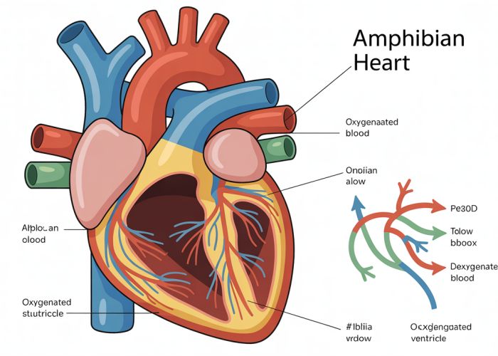
The Amphibian Heart: A Three-Chambered Wonder
Amphibians possess a three-chambered heart, a characteristic that distinguishes them from fish (typically two chambers) and most reptiles, birds, and mammals (typically four chambers). This three-chambered design presents both benefits and challenges regarding oxygenated and deoxygenated blood separation.
The Three Chambers: A Closer Examination
The amphibian heart consists of:
- Two Atria: A right atrium receiving deoxygenated blood from the body and a left atrium receiving oxygenated blood from the lungs (or gills, depending on the life stage).
- One Ventricle: This single ventricle is where both oxygenated and deoxygenated blood mix to varying degrees before being pumped out.
The Conus Arteriosus: An Extra Compartment
In some amphibians, notably some frogs and toads, a structure called the conus arteriosus is present. While not a true "chamber" in the same way as the atria and ventricle, it plays a crucial role in directing blood flow. The conus arteriosus possesses a spiral valve that assists in partially separating blood being sent to the lungs (for oxygenation) and to the rest of the body.
How it Works: The Pumping Mechanism
The amphibian heart functions through a carefully orchestrated sequence of contractions and relaxations.
- Atrial Contraction: The atria contract, simultaneously pushing blood into the single ventricle. The right atrium delivers deoxygenated blood, while the left atrium delivers oxygenated blood.
- Ventricular Contraction: The ventricle contracts, pumping blood into the arteries leading to the lungs and the rest of the body. The spiral valve (if present) assists in directing blood flow.
- Relaxation (Diastole): The heart muscles relax, allowing the atria to fill with blood again, ready for the next cycle.
The Challenge of Mixing: Minimizing Oxygenation Loss
The single ventricle presents the challenge of mixing oxygenated and deoxygenated blood. However, several mechanisms minimize this mixing and ensure that tissues receive a relatively high proportion of oxygenated blood.
- Timing of Contractions: The timing of atrial contractions and ventricular contraction helps to stratify the blood somewhat within the ventricle, reducing complete mixing.
- Spiral Valve (Conus Arteriosus): As previously mentioned, the spiral valve (if present) directs deoxygenated blood preferentially towards the pulmonary artery (to the lungs) and oxygenated blood towards the systemic arteries (to the body).
- Trabeculae: The inner walls of the ventricle contain ridges called trabeculae, which create channels that help to keep the oxygenated and deoxygenated blood partially separated.
Amphibian Heart vs. Other Vertebrate Hearts: A Comparative Analysis
Let’s compare the amphibian heart to the hearts of other vertebrates to understand its evolutionary context.
| Feature | Fish (Typical) | Amphibian | Reptile (Typical) | Bird/Mammal |
|---|---|---|---|---|
| Number of Atria | 1 | 2 | 2 | 2 |
| Number of Ventricles | 1 | 1 | 1 (partially divided) | 2 |
| Blood Mixing | Significant | Partial | Partial (less than amphibians) | Minimal/None |
| Oxygenation Efficiency | Lower | Intermediate | Intermediate to High | High |
This table highlights the evolutionary progression from a simple two-chambered heart in fish to the highly efficient four-chambered heart in birds and mammals, with the amphibian heart representing an intermediate stage. While the amphibian heart might seem less efficient than a four-chambered heart, its design is well-suited to their lifestyle, particularly their ability to breathe through their skin (cutaneous respiration), which reduces their reliance on fully oxygenated blood from the lungs.
Variation Among Amphibians: Adaptations and Specializations
While the three-chambered heart is the general rule for amphibians, variations exist that reflect specific adaptations to their environment and lifestyle.
Lungless Salamanders: A Unique Case
Lungless salamanders (Plethodontidae) have evolved to rely entirely on cutaneous respiration, lacking both lungs and gills. Their circulatory system is modified accordingly, with a reduced need for separate pulmonary and systemic circuits. While they still possess a three-chambered heart, the blood flow patterns are somewhat altered to accommodate their unique respiratory physiology. These adaptations demonstrate the remarkable plasticity and adaptability of the amphibian circulatory system.
Frequently Asked Questions About Amphibians’ Heart Chambers
Why do amphibians have a three-chambered heart?
Amphibians have evolved with a three-chambered heart as an adaptation to their lifestyle, which includes both aquatic and terrestrial environments. This heart structure allows them to efficiently manage both oxygenated and deoxygenated blood flow, despite some mixing of the blood. It’s a compromise that works for their dual-environment needs.
How does the three-chambered heart work in amphibians?
The three-chambered heart consists of two atria and one ventricle. Deoxygenated blood from the body enters the right atrium, while oxygenated blood from the lungs and skin enters the left atrium. Both empty into the single ventricle where some mixing occurs before the blood is pumped to the lungs and body.
Is the three-chambered heart as efficient as a four-chambered heart?
No, the three-chambered heart found in amphibians is generally not as efficient as the four-chambered heart seen in mammals and birds. The mixing of oxygenated and deoxygenated blood within the single ventricle reduces the overall oxygen delivery efficiency compared to systems with complete separation.
Do all amphibians have the exact same type of three-chambered heart?
While most amphibians share the basic three-chambered heart structure, there can be some variations. For instance, some amphibian species have adaptations within the ventricle to minimize the mixing of oxygenated and deoxygenated blood, improving the efficiency of their amphibians heart chambers system.
So, that’s a wrap on **amphibians heart chambers**! Hope you learned something cool and now have a better grasp of how these amazing creatures pump their blood. Keep exploring the natural world, and maybe you’ll even stumble upon a frog with a particularly impressive heart. Until next time!