Understanding the intricate mechanisms that govern hemostasis is crucial for appreciating human physiology. The complex cascade involving fibrinogen, a protein produced in the liver, is central to this process. This article will delve into the essential role of the 13 clotting factors, offering an analytical exploration of how these proteins interact to maintain vascular integrity. Leading hematology research institutions like the Mayo Clinic have dedicated significant efforts to elucidate these processes, enabling advancements in treating bleeding disorders and conditions like thrombosis. Each of the 13 clotting factors plays a unique and vital role in the coagulation process, and understanding them can unlock critical insights into human health.
Imagine a world without the ability to stop bleeding – even a minor cut could become a life-threatening event. Fortunately, our bodies possess a remarkable defense mechanism known as blood clotting, or hemostasis. This intricate process is our natural safeguard against excessive blood loss.
The Vital Role of Hemostasis
Hemostasis is far more than just a simple plug for a wound. It’s a finely tuned cascade of events designed to rapidly seal damaged blood vessels, preventing exsanguination while simultaneously initiating the healing process.
This sophisticated system relies on a team of specialized proteins called clotting factors, also known as coagulation factors.
These factors work in concert, each playing a crucial role in a carefully orchestrated sequence of reactions.
Decoding the Coagulation Factors
This article aims to demystify the world of blood clotting by exploring each of the 13 recognized clotting factors.
We will delve into their individual functions, how they interact within the coagulation cascade, and their critical importance for maintaining health.
Understanding these factors is paramount, not only for healthcare professionals but also for anyone seeking to gain deeper insight into the body’s remarkable ability to heal and protect itself.
Why Understanding Clotting Factors Matters
By exploring these factors, we can better understand the causes of bleeding disorders and thrombotic conditions.
Ultimately, knowledge of these essential proteins empowers us to promote health and prevent disease.
The Blood Coagulation Cascade: A Step-by-Step Overview
Building upon the fundamental understanding of hemostasis, it’s time to explore the core mechanism that brings it all together: the blood coagulation cascade.
This cascade is not a simple, linear process, but rather a complex, interwoven series of enzymatic reactions.
Think of it as a carefully choreographed dance, where each clotting factor activates the next in a precise order, ultimately leading to the formation of a stable blood clot.
Unveiling the Cascade: A Series of Reactions
At its heart, the coagulation cascade is a chain reaction.
One activated clotting factor acts as an enzyme, triggering the activation of the next factor in the sequence.
This process amplifies the initial signal, ensuring a rapid and effective response to vascular injury.
Each step is crucial, and a deficiency or malfunction in any one factor can disrupt the entire cascade, leading to bleeding disorders.
The Intrinsic and Extrinsic Pathways: Two Roads to a Common Goal
The coagulation cascade is traditionally divided into two main pathways: the intrinsic and the extrinsic pathways.
While they initiate clotting through different mechanisms, both pathways ultimately converge into a common pathway, culminating in the formation of fibrin, the protein that forms the meshwork of a blood clot.
The Intrinsic Pathway: Initiated Within the Blood
The intrinsic pathway is triggered by factors present within the blood itself.
It is activated when blood comes into contact with negatively charged surfaces, such as collagen exposed at the site of vessel injury.
This pathway involves factors XII, XI, IX, and VIII, along with other proteins, in a series of activation steps.
The Extrinsic Pathway: A Rapid Response
The extrinsic pathway, on the other hand, is initiated by tissue factor, a protein located outside the blood vessels.
When a blood vessel is injured, tissue factor is exposed to the blood, forming a complex with factor VII.
This complex rapidly activates factor X, initiating the common pathway.
The extrinsic pathway is generally considered to be the primary initiator of blood coagulation in response to tissue injury.
The Common Pathway: The Final Steps
Both the intrinsic and extrinsic pathways converge on the common pathway, which involves factors X, V, prothrombin (factor II), and fibrinogen (factor I).
Factor X, activated by either pathway, forms a complex with factor V.
This complex then activates prothrombin to thrombin.
Thrombin, in turn, converts fibrinogen into fibrin, the insoluble protein that forms the structural framework of the blood clot.
Finally, factor XIII, activated by thrombin, cross-links the fibrin strands, stabilizing the clot and making it resistant to breakdown.
The Indispensable Role of Clotting Factors
Within this intricate cascade, each clotting factor plays a vital and sequential role.
The absence or dysfunction of even one factor can significantly impair the body’s ability to form a stable blood clot.
This is why understanding the individual roles of these factors is so crucial for diagnosing and managing bleeding disorders.
Each factor’s precise function contributes to the overall efficiency and effectiveness of the hemostatic process.
Building upon the fundamental understanding of hemostasis, it’s time to explore the core mechanism that brings it all together: the coagulation cascade. Now, let’s transition from understanding the process as a whole to zooming in on the individual stars of the show – the thirteen clotting factors.
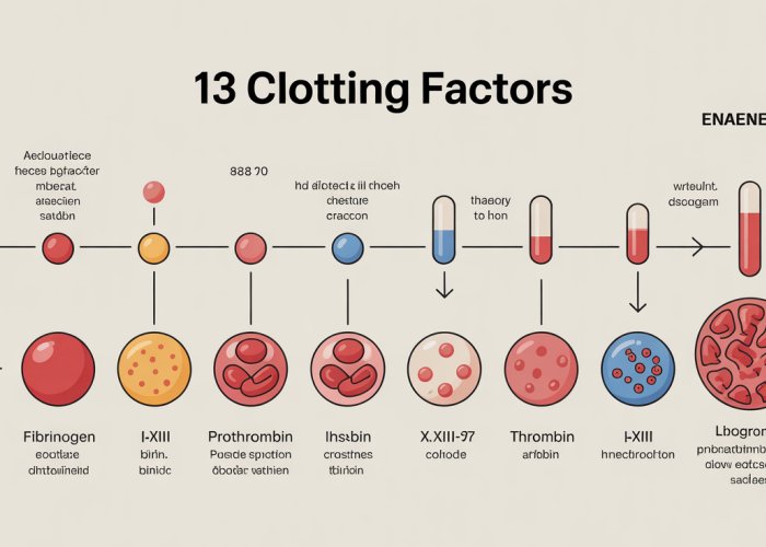
Meet the Players: A Detailed Look at the 13 Clotting Factors
Each of the thirteen recognized clotting factors plays a unique and irreplaceable role in the intricate dance of the coagulation cascade. These specialized proteins, synthesized primarily in the liver, are essential for converting liquid blood into a stable clot, effectively stopping bleeding and initiating the healing process. Let’s delve into each of these critical players, exploring their individual functions, activation mechanisms, and clinical significance.
Factor I: Fibrinogen – The Foundation of the Clot
Fibrinogen, also known as Factor I, is a large, soluble glycoprotein synthesized in the liver. Its primary role is to serve as the precursor to fibrin, the insoluble protein that forms the meshwork of a blood clot.
Thrombin, generated during the coagulation cascade, cleaves fibrinogen into fibrin monomers. These monomers then spontaneously polymerize to form fibrin strands.
Factor XIII further strengthens the fibrin meshwork. Without sufficient fibrinogen, the body’s ability to form stable clots is severely compromised.
Factor II: Prothrombin – The Thrombin Precursor
Prothrombin, or Factor II, is another vitamin K-dependent glycoprotein produced in the liver. It is the inactive precursor to thrombin, a serine protease that plays a central role in coagulation.
The activation of prothrombin to thrombin is catalyzed by the prothrombinase complex, which includes Factor Xa, Factor Va, calcium ions, and phospholipids.
Thrombin has multiple functions in the coagulation cascade, including: converting fibrinogen to fibrin, activating Factor XIII, and amplifying the activation of other coagulation factors.
Factor III: Tissue Factor – The Initiator of the Extrinsic Pathway
Tissue Factor (TF), also known as Factor III or thromboplastin, is a transmembrane protein expressed by cells outside of blood vessels, such as subendothelial fibroblasts and smooth muscle cells. TF plays a crucial role in initiating the extrinsic pathway of coagulation.
When blood vessels are injured, TF is exposed to the bloodstream. It then binds to Factor VIIa, forming the TF-VIIa complex.
This complex activates Factor X and Factor IX, initiating the coagulation cascade. The Tissue Factor pathway is considered the primary pathway for initiating blood coagulation in vivo.
Factor IV: Calcium – The Essential Cofactor
Calcium ions (Ca2+), designated as Factor IV, are essential cofactors in several steps of the coagulation cascade. Calcium ions bind to various coagulation factors and phospholipid surfaces, facilitating their interactions and promoting the assembly of enzyme complexes.
Calcium is particularly important for the activation of Factors IX, X, prothrombin, and the binding of Factors V and VIII to platelets.
The chelation (removal) of calcium ions by substances like EDTA or citrate is a common method used to prevent blood from clotting in vitro, such as in blood collection tubes for laboratory testing.
Factor V: Proaccelerin – The Prothrombinase Complex Component
Factor V, also known as proaccelerin or labile factor, is a glycoprotein that acts as a cofactor in the prothrombinase complex. This complex is responsible for converting prothrombin to thrombin.
Factor Va, the activated form of Factor V, binds to Factor Xa on the platelet surface, significantly enhancing the rate of prothrombin activation.
Factor V Leiden is a common genetic mutation that makes Factor V resistant to inactivation by activated protein C (APC), leading to an increased risk of thrombosis.
Factor VII: Proconvertin – The Extrinsic Pathway Activator
Factor VII, also known as proconvertin or stable factor, is a vitamin K-dependent serine protease. It is activated by Tissue Factor (TF) during the initiation of the extrinsic pathway.
The TF-VIIa complex activates Factor X and Factor IX, initiating the coagulation cascade.
Factor VII plays a crucial role in hemostasis, and deficiencies in Factor VII can lead to bleeding disorders.
Factor VIII: Antihemophilic Factor – The Hemophilia A Connection
Factor VIII, or antihemophilic factor, is a glycoprotein that acts as a cofactor for Factor IXa in the intrinsic pathway. It is essential for the activation of Factor X.
Factor VIII circulates in the plasma bound to von Willebrand factor (vWF), which protects it from degradation.
Deficiency or dysfunction of Factor VIII causes Hemophilia A, a severe bleeding disorder characterized by prolonged bleeding after injury or surgery. Treatment for Hemophilia A typically involves replacement therapy with recombinant Factor VIII.
Factor IX: Christmas Factor – The Hemophilia B Connection
Factor IX, also known as Christmas factor, is a vitamin K-dependent serine protease that plays a crucial role in the intrinsic pathway. It is activated by Factor XIa and, to a lesser extent, by the TF-VIIa complex.
Factor IXa, in complex with Factor VIIIa, activates Factor X. Deficiency or dysfunction of Factor IX causes Hemophilia B (Christmas disease), a bleeding disorder similar to Hemophilia A.
Treatment for Hemophilia B involves replacement therapy with recombinant Factor IX.
Factor X: Stuart-Prower Factor – The Common Pathway Convergence
Factor X, or Stuart-Prower factor, is a vitamin K-dependent serine protease that occupies a central position in the coagulation cascade. It is the point where the intrinsic and extrinsic pathways converge into the common pathway.
Factor X is activated by both the TF-VIIa complex (extrinsic pathway) and the Factor IXa-Factor VIIIa complex (intrinsic pathway).
Factor Xa, in complex with Factor Va, forms the prothrombinase complex, which activates prothrombin to thrombin.
Factor XI: Plasma Thromboplastin Antecedent – The Intrinsic Pathway Amplifier
Factor XI, also known as plasma thromboplastin antecedent (PTA), is a serine protease that plays a role in the intrinsic pathway. It is activated by Factor XIIa and thrombin.
Factor XIa activates Factor IX, which then activates Factor X. Deficiency of Factor XI leads to Hemophilia C, a milder bleeding disorder compared to Hemophilia A or B.
Factor XII: Hageman Factor – The Contact Activation Initiator
Factor XII, or Hageman factor, is a serine protease that initiates the contact activation pathway. This pathway is part of the intrinsic pathway of coagulation.
Factor XII is activated when it comes into contact with negatively charged surfaces, such as collagen, kallikrein, and high-molecular-weight kininogen (HMWK).
Activated Factor XII (XIIa) then activates Factor XI. While Factor XII initiates coagulation in vitro, its role in vivo is less clear, as individuals with Factor XII deficiency often do not have significant bleeding problems.
Factor XIII: Fibrin-Stabilizing Factor – The Clot Reinforcer
Factor XIII, also known as fibrin-stabilizing factor, is a transglutaminase that cross-links fibrin monomers, strengthening and stabilizing the fibrin clot.
Thrombin activates Factor XIII to Factor XIIIa. Factor XIIIa catalyzes the formation of covalent bonds between fibrin molecules.
This cross-linking makes the clot more resistant to degradation and enhances its mechanical strength. Factor XIII deficiency can result in delayed bleeding and poor wound healing.
Vitamin K: The Clotting Catalyst
Having explored the individual roles of the thirteen clotting factors, it’s crucial to understand the essential cofactors that enable their function. Vitamin K stands out as a critical nutrient in this respect, acting as a catalyst in the synthesis of several key players in the coagulation cascade.
This fat-soluble vitamin is indispensable for the proper production and functionality of clotting factors II (prothrombin), VII (proconvertin), IX (Christmas factor), and X (Stuart-Prower factor). Without adequate levels of Vitamin K, the liver’s ability to produce functional versions of these factors is significantly compromised, leading to potential bleeding complications.
The Vital Role of Vitamin K in Clotting Factor Synthesis
Vitamin K’s role in the synthesis of clotting factors is linked to a crucial post-translational modification process. This modification, known as gamma-carboxylation, is essential for these factors to bind calcium ions (Factor IV), enabling their interaction with phospholipid surfaces in the coagulation cascade.
Specifically, Vitamin K acts as a cofactor for the enzyme gamma-glutamyl carboxylase.
This enzyme adds a carboxyl group to specific glutamic acid residues on the precursor proteins of factors II, VII, IX, and X. This carboxylation is absolutely essential for the proper function of these clotting factors.
Without this modification, the clotting factors are unable to participate effectively in the coagulation cascade, hindering the body’s ability to form blood clots properly.
Vitamin K Deficiency: Consequences and Causes
Vitamin K deficiency can arise from various factors, including inadequate dietary intake, malabsorption issues, and certain medications.
Newborn infants are particularly vulnerable to Vitamin K deficiency bleeding (VKDB) due to limited placental transfer of Vitamin K and low levels in breast milk, and immature gut flora.
Adults who suffer from conditions that impair nutrient absorption, such as cystic fibrosis, celiac disease, or short bowel syndrome, may also develop Vitamin K deficiencies. Certain medications, such as warfarin (a vitamin K antagonist), can also induce Vitamin K deficiency and increase bleeding risk.
Bleeding Disorders and Vitamin K Deficiency
The clinical consequence of Vitamin K deficiency is an increased risk of bleeding. This can manifest as easy bruising, nosebleeds, gum bleeding, and, in severe cases, life-threatening hemorrhages.
Newborns with VKDB may experience intracranial hemorrhage, which can lead to serious neurological damage.
Therefore, ensuring adequate Vitamin K levels, through diet or supplementation, is crucial for maintaining healthy blood clotting function and preventing bleeding complications. The recommended daily intake varies depending on age and individual health status. It’s often prudent to consult with a healthcare professional to determine optimal Vitamin K intake, especially for individuals with pre-existing conditions or those taking medications that may interfere with Vitamin K metabolism.
When Things Go Wrong: Clotting Factor Deficiencies, Bleeding Disorders, and Thrombosis
While a functional coagulation cascade diligently protects us from excessive bleeding, its disruption can lead to serious health issues. Deficiencies in clotting factors, whether inherited or acquired, manifest as bleeding disorders, impairing the body’s ability to form clots properly. Understanding these disorders is critical for diagnosis, management, and improving patient outcomes. However, the opposite extreme – inappropriate clotting – also poses significant risks, highlighting the delicate balance within the hemostatic system.
Common Bleeding Disorders: A Closer Look
Several well-defined bleeding disorders are directly linked to specific clotting factor deficiencies. Here’s a concise overview of some of the most prevalent:
-
Hemophilia A (Factor VIII Deficiency):
This is a classic example of a bleeding disorder caused by a deficiency in Factor VIII. Hemophilia A is a genetic disorder, typically inherited in an X-linked recessive pattern, primarily affecting males. The severity of the condition varies, ranging from mild to severe, depending on the level of functional Factor VIII present in the blood. Clinical presentation often involves prolonged bleeding after injuries or surgery, spontaneous bleeding into joints (hemarthrosis), and easy bruising. -
Hemophilia B (Factor IX Deficiency):
Also known as Christmas disease, Hemophilia B stems from a deficiency in Factor IX. Similar to Hemophilia A, it is also an X-linked recessive disorder, predominantly affecting males. Its clinical manifestations are nearly identical to those of Hemophilia A, making laboratory testing crucial for accurate diagnosis and differentiation. -
Von Willebrand Disease (vWD):
Unlike Hemophilia A and B, Von Willebrand disease is usually an autosomal disorder, meaning it affects both males and females. It is characterized by a deficiency or dysfunction of von Willebrand factor (vWF), a protein that helps platelets adhere to the site of injury and carries Factor VIII in the bloodstream. vWD is the most common inherited bleeding disorder, with varying degrees of severity. Symptoms can include easy bruising, nosebleeds, heavy menstrual bleeding (menorrhagia) in women, and prolonged bleeding after dental procedures or surgery.
Genetic Basis and Clinical Presentation
The genetic basis of these bleeding disorders plays a crucial role in their inheritance patterns and clinical presentation.
For instance, the X-linked recessive inheritance of Hemophilia A and B explains why males are predominantly affected, as they only have one X chromosome.
Females, possessing two X chromosomes, usually act as carriers, potentially passing the affected gene to their offspring. The severity of symptoms can also vary significantly, depending on the specific genetic mutation and its impact on the production or function of the affected clotting factor.
The Other Side of the Coin: Thrombosis
While deficiencies in clotting factors lead to bleeding disorders, excessive or inappropriate activation of the coagulation cascade can result in thrombosis, the formation of blood clots within blood vessels. Thrombosis can obstruct blood flow, leading to severe complications such as deep vein thrombosis (DVT), pulmonary embolism (PE), stroke, and heart attack.
Understanding the intricacies of the clotting cascade is paramount in preventing and treating thrombotic events.
Anticoagulant medications, such as warfarin and heparin, target specific clotting factors or pathways within the cascade, effectively reducing the risk of clot formation. Moreover, identifying individuals at high risk for thrombosis, such as those with inherited thrombophilia (a predisposition to clotting) or acquired risk factors like prolonged immobility or surgery, allows for proactive preventative measures.
The Liver’s Contribution: Manufacturing Clotting Factors
The intricate dance of the coagulation cascade relies heavily on the liver, a metabolic powerhouse responsible for synthesizing the majority of clotting factors circulating in our bloodstream. Its role in maintaining hemostasis is so profound that liver dysfunction can rapidly translate into significant bleeding complications. This section delves into the liver’s crucial contribution to blood clotting and explores the consequences of impaired liver function on the coagulation system.
The Liver: A Clotting Factor Factory
The liver is the primary site of synthesis for several key clotting factors, including:
- Factor I (Fibrinogen)
- Factor II (Prothrombin)
- Factor V (Proaccelerin)
- Factor VII (Proconvertin)
- Factor IX (Christmas Factor)
- Factor X (Stuart-Prower Factor)
- Protein C
- Protein S
- Antithrombin
These factors are essential components of both the intrinsic and extrinsic pathways of the coagulation cascade, making the liver’s synthetic capacity indispensable for normal blood clotting. Without sufficient production of these factors, the body’s ability to form stable clots is severely compromised.
Vitamin K-dependent clotting factors (II, VII, IX, and X) undergo a post-translational modification in the liver. This modification, carboxylation, is essential for their function. Without Vitamin K, these factors cannot bind calcium, preventing their participation in the coagulation cascade.
Liver Disease: A Threat to Hemostasis
Liver diseases, such as cirrhosis, hepatitis, and liver failure, significantly impair the liver’s ability to synthesize clotting factors. This reduced production leads to a deficiency in these vital proteins, disrupting the coagulation cascade and increasing the risk of bleeding.
The severity of the bleeding risk often correlates with the extent of liver damage. Advanced liver disease can result in critically low levels of multiple clotting factors, leading to spontaneous bleeding, prolonged bleeding after minor injuries, and an increased risk of hemorrhage during surgery or other invasive procedures.
The Impact of Liver Dysfunction on Coagulation
Beyond the reduced synthesis of clotting factors, liver disease impacts hemostasis in several other ways:
-
Reduced Clearance of Activated Clotting Factors: The liver normally clears activated clotting factors from the circulation, preventing uncontrolled clot formation. Liver dysfunction impairs this clearance, potentially leading to a paradoxical situation where both bleeding and thrombotic risks are elevated.
-
Thrombocytopenia: Liver disease, especially cirrhosis, can lead to thrombocytopenia (low platelet count) due to splenic sequestration of platelets or decreased thrombopoietin production, further contributing to bleeding risk.
-
Increased Fibrinolysis: The liver also plays a role in regulating fibrinolysis, the process of breaking down blood clots. Liver disease can disrupt this balance, leading to increased fibrinolysis and a predisposition to bleeding.
-
Vitamin K Malabsorption: Cholestatic liver diseases interfere with bile secretion, which is necessary for the absorption of fat-soluble vitamins like Vitamin K. This malabsorption can exacerbate deficiencies in Vitamin K-dependent clotting factors.
Assessing Coagulation in Liver Disease
Evaluating coagulation function is crucial in patients with liver disease. Traditional coagulation tests like Prothrombin Time (PT) and Partial Thromboplastin Time (PTT) are commonly used to assess clotting factor deficiencies. However, these tests may not fully reflect the complex hemostatic abnormalities in liver disease.
Viscoelastic hemostatic assays (VHA) like thromboelastography (TEG) and rotational thromboelastometry (ROTEM) offer a more comprehensive assessment of coagulation in liver disease. These tests evaluate the overall clot formation process, including clot strength and stability, providing valuable information for guiding treatment decisions.
Managing Bleeding Risk in Liver Disease
Managing bleeding risk in patients with liver disease requires a multifaceted approach:
-
Treating the Underlying Liver Disease: Addressing the underlying cause of liver disease is paramount to improving liver function and, consequently, clotting factor synthesis.
-
Vitamin K Supplementation: Administering Vitamin K can improve clotting factor synthesis in patients with Vitamin K deficiency, especially those with cholestatic liver disease.
-
Transfusion of Blood Products: In cases of severe bleeding or before invasive procedures, transfusions of fresh frozen plasma (FFP) or cryoprecipitate can provide clotting factors and fibrinogen, respectively.
-
Recombinant Activated Factor VIIa (rFVIIa): Although controversial, rFVIIa can be used as a temporary hemostatic agent in severe bleeding unresponsive to other treatments.
-
Thrombopoietin Receptor Agonists (TPO-RAs): In patients with cirrhosis and thrombocytopenia, TPO-RAs can increase platelet counts and reduce bleeding risk.
The liver’s role in synthesizing clotting factors is undeniable, making it a cornerstone of the hemostatic system. Understanding the impact of liver disease on coagulation is essential for effectively managing bleeding complications and improving patient outcomes. Recognizing the multifaceted ways in which liver dysfunction disrupts hemostasis allows clinicians to tailor treatment strategies and minimize the risk of life-threatening bleeding events.
Assessing Clotting Function: Common Diagnostic Tests
The liver’s synthetic capacity, as we’ve explored, is indispensable for producing the clotting factors essential for hemostasis. But how do clinicians assess whether these factors are functioning correctly and in sufficient quantities? The answer lies in a series of diagnostic tests designed to evaluate the intricate processes of the coagulation cascade.
Prothrombin Time (PT): Evaluating the Extrinsic Pathway
Prothrombin Time (PT) is a blood test that measures how long it takes for a clot to form in a sample of plasma after the addition of thromboplastin, a reagent that initiates the extrinsic coagulation pathway. This test primarily assesses the function of factors I (fibrinogen), II (prothrombin), V, VII, and X.
The PT test is a valuable tool for evaluating:
- Liver function.
- Vitamin K status.
- Warfarin (Coumadin) therapy.
A prolonged PT indicates that one or more of these factors are deficient or not functioning correctly, potentially due to liver disease, vitamin K deficiency, or the effects of anticoagulant medications like warfarin.
Partial Thromboplastin Time (PTT): Evaluating the Intrinsic Pathway
Partial Thromboplastin Time (PTT) measures the time it takes for a clot to form in a plasma sample after the addition of a contact activator (such as silica or kaolin) and phospholipids. This test evaluates the function of the intrinsic and common coagulation pathways, assessing factors I (fibrinogen), II (prothrombin), V, VIII, IX, X, XI, and XII.
The PTT test is particularly useful for:
- Detecting Hemophilia A (Factor VIII deficiency) and Hemophilia B (Factor IX deficiency).
- Monitoring heparin therapy.
- Identifying lupus anticoagulants.
A prolonged PTT suggests a deficiency or dysfunction in one or more of these factors, potentially due to genetic disorders like hemophilia or the presence of inhibitors that interfere with the coagulation cascade.
Interpreting PT and PTT Results: A Clinical Puzzle
Interpreting PT and PTT results requires careful consideration of the patient’s clinical history, medication use, and other laboratory findings. A prolonged PT or PTT can indicate:
- Clotting factor deficiencies: Congenital or acquired.
- Vitamin K deficiency: Impairing the function of factors II, VII, IX, and X.
- Liver disease: Reducing the synthesis of clotting factors.
- Anticoagulant therapy: Warfarin and heparin prolong PT and PTT, respectively.
- Presence of inhibitors: Such as lupus anticoagulants.
Further investigations, such as individual factor assays, may be necessary to pinpoint the specific cause of the abnormality.
International Normalized Ratio (INR): Standardizing PT Results
The International Normalized Ratio (INR) is a calculation based on the PT result that standardizes the measurement across different laboratories using different thromboplastin reagents. The INR is calculated using the following formula:
INR = (PTpatient / PTnormal)ISI
Where:
- PTpatient is the patient’s prothrombin time.
- PTnormal is the mean normal prothrombin time for the laboratory.
- ISI is the International Sensitivity Index, a value assigned to each thromboplastin reagent that indicates its sensitivity to vitamin K-dependent clotting factors.
The INR is particularly important for monitoring warfarin therapy, as it provides a consistent and reliable measure of the drug’s effect on coagulation, regardless of the laboratory performing the test. A target INR range of 2.0 to 3.0 is typically desired for patients taking warfarin for conditions such as atrial fibrillation or venous thromboembolism, but this range may vary depending on the indication and individual patient characteristics.
The Broader Picture
While PT and PTT are essential screening tests, they represent only a snapshot of the complex coagulation system. Other tests, such as fibrinogen levels, D-dimer assays, and specific factor assays, may be necessary to fully evaluate a patient’s clotting function and guide appropriate management.
Partial Thromboplastin Time (PTT) offers vital insights into the intrinsic and common pathways, complementing the information gleaned from Prothrombin Time (PT). Together, these tests provide a comprehensive overview of a patient’s clotting abilities, guiding clinical decisions regarding diagnosis and treatment. But what happens when the body’s own coagulation mechanisms need assistance, when the risk of excessive clotting looms large?
Manipulating the Cascade: Medications That Affect Clotting
Sometimes, the body’s natural clotting mechanisms can become overactive, leading to the formation of dangerous blood clots. These clots, if left unchecked, can cause strokes, heart attacks, or other life-threatening conditions. Fortunately, medical science offers a range of medications designed to interfere with the coagulation cascade, effectively reducing the risk of clot formation. These medications, known as anticoagulants, play a crucial role in preventing and treating thromboembolic disorders.
Warfarin: Vitamin K Antagonist
Warfarin (Coumadin) stands as one of the most widely used oral anticoagulants. Its mechanism of action centers on disrupting the Vitamin K cycle, which is essential for the synthesis of several crucial clotting factors: II, VII, IX, and X.
By interfering with Vitamin K-dependent carboxylation, Warfarin effectively reduces the production of these factors, leading to a decrease in the blood’s ability to clot.
Clinical Uses and Monitoring
Warfarin is commonly prescribed for long-term anticoagulation in conditions such as:
- Atrial fibrillation (to prevent stroke).
- Venous thromboembolism (deep vein thrombosis and pulmonary embolism).
- Mechanical heart valves.
Because Warfarin’s effectiveness can be affected by diet, other medications, and individual patient factors, careful monitoring is crucial.
The International Normalized Ratio (INR), derived from the Prothrombin Time (PT) test, is used to standardize Warfarin dosage and ensure that patients remain within a safe and therapeutic range. Regular blood tests are essential to maintain optimal anticoagulation.
Heparin: Activating Antithrombin
Heparin, in contrast to Warfarin, acts more directly and rapidly on the coagulation cascade. It’s typically administered intravenously or subcutaneously.
Heparin’s primary mechanism involves enhancing the activity of antithrombin, a naturally occurring anticoagulant protein in the blood. Antithrombin inhibits several clotting factors, including thrombin (Factor IIa) and Factor Xa.
By boosting antithrombin’s activity, Heparin effectively shuts down multiple steps in the coagulation cascade, preventing further clot formation.
Unfractionated Heparin (UFH) vs. Low-Molecular-Weight Heparin (LMWH)
Heparin exists in two primary forms:
-
Unfractionated Heparin (UFH): Requires close monitoring of aPTT (activated partial thromboplastin time) due to its variable effects.
-
Low-Molecular-Weight Heparins (LMWH): Such as enoxaparin (Lovenox) and dalteparin (Fragmin), offer more predictable responses and can often be administered without intensive lab monitoring.
Clinical Applications
Heparin is used in a variety of clinical settings, including:
- Acute treatment of venous thromboembolism.
- Prevention of clotting during surgery or dialysis.
- Management of acute coronary syndromes (e.g., unstable angina, myocardial infarction).
Direct Oral Anticoagulants (DOACs): A Newer Generation
A newer class of anticoagulant medications, known as Direct Oral Anticoagulants (DOACs), has emerged in recent years. These drugs offer several advantages over Warfarin, including more predictable dosing and reduced need for routine monitoring.
DOACs directly inhibit specific clotting factors, such as:
- Thrombin (Factor IIa): Dabigatran (Pradaxa).
- Factor Xa: Rivaroxaban (Xarelto), Apixaban (Eliquis), and Edoxaban (Savaysa).
Benefits and Considerations
DOACs have proven effective in preventing stroke in patients with atrial fibrillation and treating/preventing venous thromboembolism. Their ease of use and reduced monitoring requirements have made them increasingly popular.
However, it’s essential to consider potential risks, such as bleeding, and to carefully assess individual patient factors before prescribing these medications.
Balancing the Risks
Anticoagulant medications are powerful tools in managing clotting disorders, but they also carry the risk of bleeding. Therefore, careful patient selection, appropriate dosing, and ongoing monitoring are crucial to maximize benefits and minimize potential harm.
FAQs: Decoding the 13 Clotting Factors
Here are some common questions to help you further understand the role and importance of the 13 clotting factors in blood coagulation.
What happens if one of my 13 clotting factors is deficient?
A deficiency in one of the 13 clotting factors can disrupt the normal blood clotting process. This can lead to prolonged bleeding after injury, easy bruising, or in severe cases, internal bleeding. The specific symptoms and severity depend on which factor is deficient and the extent of the deficiency.
How do the 13 clotting factors work together to stop bleeding?
The 13 clotting factors work in a complex cascade, where each factor activates the next in a specific sequence. This cascade ultimately results in the formation of a stable blood clot, which stops the bleeding. Think of it like a domino effect, where each factor plays a vital role.
Can I increase my levels of the 13 clotting factors if they are low?
Treatment for low levels of the 13 clotting factors depends on the specific factor that’s deficient and the underlying cause. Options may include infusions of clotting factor concentrates, medications, or addressing underlying medical conditions. Consultation with a hematologist is crucial.
Are there any genetic conditions related to the 13 clotting factors?
Yes, several inherited bleeding disorders are caused by genetic mutations affecting the 13 clotting factors. Hemophilia A and B, for example, are caused by deficiencies in factors VIII and IX respectively. These conditions are typically diagnosed early in life due to prolonged bleeding episodes.
So, that’s the lowdown on the 13 clotting factors! Hopefully, you found that helpful and can now impress your friends with your newfound knowledge. Stay healthy!